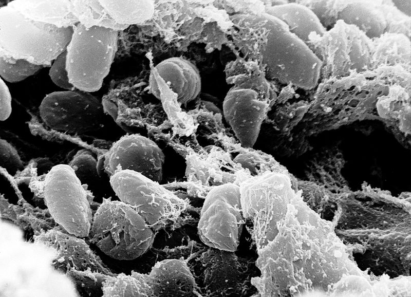
Yersinia are short, encapsulated, non motile gram negative rods.
Transmission:
Humans are the accidental hosts (bitten by a flea that is a part of sylvatic cycle). Sylvatic cycle consists of transmission among mild rodents by flea. Transmission also occurs by respiratory droplets from infected patients.
Pathogenesis
Rodents and flies are the selective hosts, the events leading to human beings biting are:
1. Flea ingests the infected bacteria from infected rodent
2. As a result of coagulase, the blood clots in fleas stomach
3. The bacteria are trapped in fibrin
4. Bacteria proliferate to large numbers
5. The mass of bacteria and fibrin blocks the proventriculus of fleas intestine
6. On next feeding flea regurgitates the organism into animals
7. Because proventriculus is blocked and flea gets no nutrition, so is hungry and loses its host selectivity
8. Pathogen breaches the skin through dermal injuries
9. Bacteria reaches regional lymph nodes
In 2 to 5 days after infection, blue and swollen lymph nodes are observed. Bacteria break into blood stream to cause sepsis. Dissemination into pulmonary circulation results in secondary pulmonary plaque.
Capsule, called F1, protects against phagocytosis. Bacteria releas an endotoxin, the exotoxic reaction is unknown. 2 proteins V antigen and W antigen allow the organism to survive and grow intracellularly.
Yersinia outer protein called ‘yops’ is injected into humans via type 3 secretion system and inhibits phagocytosis and cytokine production by macrophages and neutrophils.
Virulence factors:
• Capsule F1
• Endotoxin
• Exotoxin
• V antigen
• W antigen
• Yersinia outer proteins
Clinical symptoms:
• Pain and swollen lymph nodes
• High fever
• Septic shock
• Pneumonia
• Myalgias
• Prostration
Lab diagnosis:
Specimen
• Blood
• Pus
• Buboaspirates (care needed when handling)
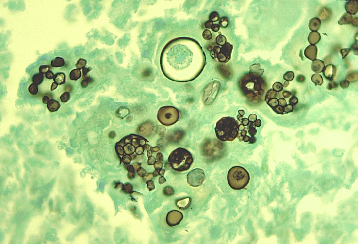
Microscopy:
• Gram negative rods
• Encapsulated
• Short
• Non motile
• Bipolar staining, resemble a safety pin with a central clear area
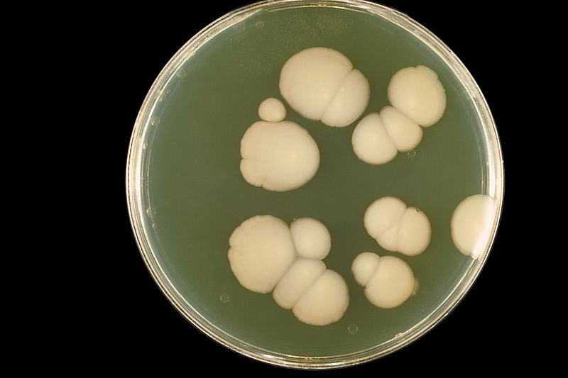
Culture:
Blood Agar
On blood agar, small, shiny non hemolytic colonies are formed (after 24 to 48 hours of incubation)
MacConkey’s agar
On MacConkey’s agar, very small, translucent, pink colonies are produced (incubated for 24 to 48 hours)
Giemsa and Wayson stains may be applied as well. Safety pin appearance is observed.
Biochemical Tests:
• Catalase positive
• Oxidase negative
Serological tests:
• Fuorescent – antibody staining
• Dip stick test
• PCR
Treatment:
Combination of tetracycline and tetromycin is used.