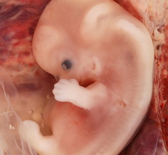
1) Gastrulation
•It is the formative process by which three germ layers, which are precursors of all embryonic tissues, and axial orientation are established in embryo.
- Ectoderm
- Mesoderm
- Endoderm
- Bilaminar germ disc converts into trilaminar germ disc.
- Embryo is referred as gastrula.
- Appearance of primitive streak.
- Formation of three germ layers.
HUMAN GASTRULATION:
Ist sign-Formation of primitive streak on the surface of epiblast
Is an opacity formed by thickened linear band of epiblast, appears caudally in the median plane of dorsal aspect of embryonic disc
Results from proliferation and movement of cells of epiblast to median plane of embryonic disc. Streak elongates by addition of cells at its caudal end. Cranial end proliferates to form Primitive node. Concurrently a narrow groove develops in the sheet that is continuous with a small depression in the primitive node called primitive pit.
Upon arrival in the region of streak, epiblast cells become flask shaped, detach from epiblast, slip beneath it. This inward movement is called invagination.
Once the cells invaginate through the groove some displace the hypoblast and forms embryonic endoderm. Others lie between epiblast and newly formed endoderm and form mesoderm. Cells remaining in the epiblast form the embryonic ectoderm.
As more and more cells move between epiblast and hypoblast they begin to spread laterally & cranially.
Gradually migrate beyond the margins of the disc and establish contact with extraembryonic mesoderm.
In cephalic direction they pass on each side of Prechordal plate.
PRECHORDAL PLATE
•Forms between the tip of notochord and buccopharyhgeal membrane and is derived from some of the first cells that migrate from the node.
•Lies b/w tip of notochord and buccopharyngeal membrane. Later prechordal plate is important for the induction of forebrain.
•Buccopharyngeal membrane:
lies at the cranial end of disc & consist of tightly adherent ectoderm & endoderm cells and represent future opening of oral cavity.
FATE OF PRIMITIVE STREAK
•Actually forms mesoderm until the end of 4th week and then regress & degenerate and soon disappears.
Remnant of primitive streak
•Sacrococcygeal teratoma —- clusters of pluripotent cells and contain tissues derived from all the three germ layers.
•Most common tumor in newborn : 1 in 37000
THUS THE EPIBLAST THROUGH THE PROCESS OF GASTRULATION IS THE SOURCE OF ALL OF THE GERM LAYERS AND CELLS IN THESE LAYERS WILL GIVE RISE TO ALL OF THE TISSUES AND ORGANS IN THE EMBRYO
2) NOTOCHORD FORMATION
Prenotochordal cells invaginating in the primitive pit move forward cranially until they reach the prechordal plate, forming a median cellular cord, the notochordal process.
Formation of notochordal canal
The floor of the notochordal process fuses with the underlying embryonic endoderm.
The fused layers gradually undergo degeneration, resulting in the formation of openings in the floor of the notochordal process, which brings the notochordal canal into communication with the yolk sac.
The openings rapidly become confluent and the floor of the notochordal canal disappears; the remains of the notochordal process form a flattened, grooved notochordal plate.
Beginning at the cranial end of the embryo, the notochordal cells proliferate and the notochordal plate infolds to form the notochord.
The proximal part of the notochordal canal persists temporarily as the neurenteric canal , which forms a transitory communication between the amniotic and yolk sac cavities.
When development of the notochord is complete, the neurenteric canal normally obliterates.
The notochord becomes detached from the endoderm of the yolk sac, which again becomes a continuous layer and itself forms the definitive notochord.
Importance:
- Defines the primordial longitudinal axis of the embryo and gives it some rigidity.
- Provides signals that are necessary for the development of axial musculoskeletal structures and the central nervous system.
- Contributes to the intervertebral discs.
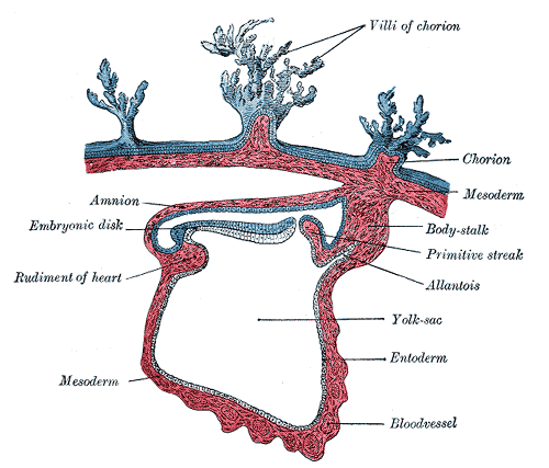
ALLANTOIS FORMATION (sausage shaped diverticulum)
16th day
In humans allantoic sac remains small, but allantoic mesoderm forms blood vessels that serve placenta and later become umblical arteries.
•The proximal part of original allantoic diverticulum persist as urachus which extends from bladder to umblicus. In adults represented by median umblical ligament

o Neural plate and neural tube formation
o Neural crest formation
Neural Crest cells (give rise to)
n Spinal ganglia (dorsal root ganglia).
n Autonomic nervous system ganglia.
n Ganglia of cranial nerves V, VII, IX, X (partly derived).
n Neurolemmal sheath of peripheral vessels.
n (Also contributes to) formation of pigment cells, supra renal medulla and various connective tissue components in the head.
4) DEVELOPMENT OF SOMITES
5) DEVELOPMENT OF INTRAEMBRYONIC COLEOM
6) EARLY DEVELOPMENT OF CARDIOVASCULAR SYSTEM
7) DEVELOPMENT OF CHORIONIC VILLI
Remnants of notochordal tissue
- CHORDOMAS: the tumours developing from remnants of notochordal tissue.
- Can be benign and malignant.
- Approx. 1/3 occur at the base of cranium and extend to the nasopharynx.
- Slowly growing.
- Malignant forms infiltrate bone.
Clinical Correlates
1) Teratogenesis associated with gastrulation:
a. Sirenomelia
b. Situs inversus
2) Tumours associated with gastrulation:
Sacrococcygeal teratomas
GROWTH OF THE EMBRYONIC DISC
•Initially flat & round, gradually becomes elongated with broad cephalic and narrow caudal end.
•In cephalic part germ cells begin their differentiation while the caudal part differentiates by the end 4th week.
•Thus while gastrulation is occuring in caudal segments cranial structures are differentiating—— cephalocaudally
Further development of trophoblast
- Primary Villus
- Secondary Villus
- Tertiary Villus
- Stem or anchoring Villus
- Free (terminal Villus)
Capillaries of tertiary villi establish contact with capillaries in mesoderm of chorionic plate and connecting stalk, inturn contact with intraembryonic circulation
TROPHOBLAST AT THE END OF THIRD WEEK OF DEVELOPMENT
- Heart begins to beat in the 4th week of development.
- Radial appearance
- Intervillous spaces lined with syncytiotrophoblast
- Outer cytotrophoblast shell

It really makes sense. Short and precise.
Thank you. made more sense
nice
Thanks a lot