Human Embryonic Period (3rd to 8th Week)
- This is the organogenetic period in which all major external and internal structures are established and the main organ systems have begun to develop.
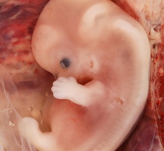
- By the 8th week the embryo has a distinctly human appearance.
- Referred as neurula
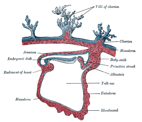
Derivatives of the Ectodermal Germ Layer:
- Disc in the 3RD Week:
The shape of the disc is from egg shape to pear shape and later becomes slipper shaped.
- Induction by notochord and prechordal mesoderm takes place.
Neurulation:
- The processes involved in the formation of the neural plate, neural folds & closure of folds to form neural tube are known as neurulation.
- Completes by the end of 4th week.
- Cells of the plate form the neuroectoderm
After induction, the neural plate gradually expands towards the primitive streak.
On about 18th day neural plate invaginates along its central axis.
The lateral edges of neural plate become more elevated to form neural folds and the depressed mid region forms the neural groove.
Gradually the neural folds fuse
The folding begins in the cervical region (5th somite)
Neural plate is converted into neural tube
The neural tube communicates with amniotic cavity.
This process completes by the end of 4th week.
After Neurulation:
The neural tube soon separates from surface ectoderm and the free edges of the surface ectoderm fuse so that this layer continues over the neural tube and the back of embryo.
Surface ectoderm differentiates into epidermis.
CNS is represented by a closed tubular structure, in which the spinal cord is locaed caudally & brain vesicles cranially.
Neural Crest Cells:
Cells at the lateral border of neuroectoderm dissociate as the neural folds elevate and fuse.
These cells undergo an epithelial – mesenchymal transition and leave the neuroectoderm to enter the underlying mesoderm.
INDUCTION OF NEURAL CREST CELLS requires an interaction between adjacent neural and overlying ectoderm.
Fate of Crest Cells from the Trunk Region:
The crest cells migrate along one of the TWO PATHWAYS:
A dorsal pathway through the dermis where they will enter the ectoderm through holes in the basal lamina to form melanocytes in the skin and hair follicles.
A ventral pathway through anterior half of each somite to become sensory ganglia, sympathetic and enteric neurons, schwann cells and cells of adrenal medulla.
Fate of the Crest Cells from Cranial Neural Folds:
LEAVE THE NEURAL FOLDS BEFORE CLOSURE
These cells contribute to the:
- Craniofacial skeleton
- Neurons for cranial ganglia, glial cells, melanocytes and other cell types
Neural Crest Derivatives:
- CONNECTIVE TISSUE & BONES OF FACE & SKULL
- CRANIAL NERVE GANGLIA
- C CELLS OF THYROID GLAND
- CONOTRUNCAL SEPTUM IN THE HEART
- ODONTOBLAST
- DERMIS IN FACE & NECK
- SPINAL (DORSAL ROOT) GANGLIA
- SYMPATHETIC CHAIN & PREAORTIC GANGLIA
- PARASYMPATHATIC GANGLIA OF GIT
- ADRENAL MEDULLA
- SCHWANN CELLS }GLIAL CELLS
- ARACHNOID & PIA MATER
- MELONOCYTES
Bilateral Ectodermal Thickenings:
- Otic placodes
- Lens placodes
Derivatives of the Ectodermal Germ Layer:
- Central Nervous System
- Peripheral Nervous System
- Sensory Epithelium of ear, nose and eye
- Epidermis including hair and nails
- S/C glands, mammary glands, pituitary gland and enamel of teeth
Abnormal Neurulation:
Neural tube defects —- 16 per 10,000 births }The two most common neural tube defects are spina bifida and anencephaly.
Anencephaly —- partial absence of brain due to failure of neural folds to fuse.
Spina bifida means cleft spine, which is an incomplete closure in the spinal column. There is usually nerve damage that causes at least some paralysis of the legs.
Three types:
- Spina Bifida Occulta
- Meningocele
- Myelomeningocele
Want a clearer concept, also see
Derivatives of Mesoderm (Embryonic Period)

the work a litle bit understanding,a big thank to the author of this work