Case Scenario:
A 15 year old[1] boy was hit on the temple[2a][2b] with a baseball and he became unconscious[3]. After about ten minutes, he regained consciousness, but he soon became lethargic, and over the next two hours, he was stuporous[4]. His pupils were unequal[5]. Intra cranial hemorrhage was suspected. He was shifted to the radiology department for an urgent CT scan[6], which confirmed a left sided epidural hematoma[7].
Explanation:
[1] Age:
As a person ages, the dura becomes more adherent to the skull, which reduces the frequency of epidural hematoma.
[2a] Region of Temple:
Temple is the side of the head behind the eye. The bones involved are the temporal mainly, along with a small part of the sphenoid.
[2b] Incidence:
The temporal region is most frequently involved in epidural hematoma (70-80% of the cases) as the bone is thin and the middle meningeal artery lies close to it.
[3] Fainting:
The state of being unconscious in EDH depends on the force of impact. The patient might undergo no loss of consciousness, brief loss or a prolonged one. Initially the force which causes the head injury is the cause of alteration of consciousness. After the patient recovers, the epidural hematoma continues to expand, increasing the intracranial pressure which might lead to a decreased level of consciousness.
[4] Stuporous:
State of mental numbness.
Lethargy is due to inadequate blood supply to the brain as well as its loss, which contributes to hypertension. In certain cases of intracranial hypertension it might lead to Cushing triad involving respiratory depression, systemic hypertension and bradycardia.
[5] Anisocoria:
Unequal pupil size. Pupil is the black part in the center of the eye which becomes larger in dim light.
The possible causes of unequal pupil sizes are varied . Some are given below:
1. Aneurysm
2. Bleeding inside cranial cavity
3. Brain tumour
4. Excess pressure due to glaucoma
5. Meningitis
6. Migraine headache
7. Use of eye drops
[7] Epidural/ Extradural Hematoma:
It occurs in the potential space between the dura and the cranium and results from the interruption of dural vessels including branches of the middle meningeal artery, veins, dural venous sinuses and skull vessels.
Etiology:
a. Trauma- most important cause
b. Infectious disease
c. Vascular malformation of the dura
d. Metastasis to the skull
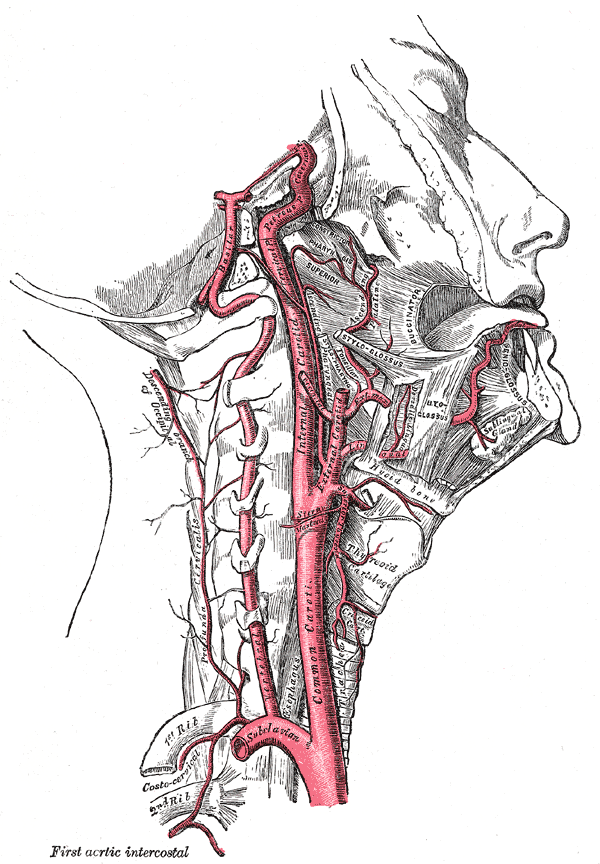
Middle meningeal artery:
The anterior division of the middle meningeal artery is most commonly involved.
Foramen spinosum, located in the middle cranial fossa, transmits the middle meningeal artery from the infratemporal fossa into the cranial cavity. The artery runs forward and laterally in a groove on upper surface of squamous part of temporal bone and the greater wing of sphenoid. After a short distance, the artery divides into anterior and posterior divisions. The anterior branch passes forward and upward to the anteroinferior angle of the parietal bone. Here the bone is deeply grooved by the artery for a short distance and is the site of damage after a blow. Artery then runs backwards and upwards on the parietal bone.
Frequency:
Epidural hematoma occurs in 10-20% of the head injuries.
Laboratory investigations:
Lab investigations involve measuring the following:
1. Hematocrit levels
2. Coagulation profile
3. Tissue thromboplastin levels
Imaging:
a. Radiograph
[6] b. CT scan:
The space occupied by epidural hematoma is limited by adherence of dura to the inner table of the skull, especially at the sutural lines, which contributes to the lenticular or biconvex appearance in the CT scan. The hematoma is denser and homogenous so easily identified.
c. MRI
Treatment:
1) Immediate surgical intervention- ligation or plugging
2) Conservative close cranial observation
The treatment is in accordance with the condition of the patient at the time of presentation.


E-medicine gives two statements…
“Association of hematoma and skull fracture is less common in young children because of calvarial plasticity.”
isn’t it contradictory to….?
“EDH is uncommon in elderly patients because the dura is strongly adhered to the inner table of the skull.”
I believe both statements are correct in their own respects. Although mortality rate is higher both in younger(less than 5 years) and older age(greater than 55 years) but the incidence is less. Most vulnerable age is somewhere less than 20 ( more than 60 %) as indicated in the article you have mentioned.