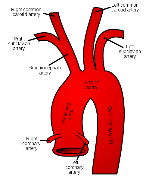
The right and left subclavian arteries have separate origins. The right subclavian artery is one of the two terminal branches of the brachiocephalic artery, the other being the right common carotid artery. It arises at the level of the right sternoclavicular joint. It moves as a gentle curve behind the scalenus anterior muscle passing upwards and laterally. At the outer border of the first rib, it becomes the axillary artery.
The left subclavian artery arises behind the left common carotid artery, from the arch of aorta. It arches in a manner similar to the right subclavian artery after reaching the root of the neck.
Grey’s Anatomy 20th edition
The scalenus anterior muscle divides the subclavian artery into three parts:
First part of the Subclavian Artery:
The first part of the subclavian artery begins from the origin of subclavian artery and extends to the medial border of the scalenus anterior muscle.
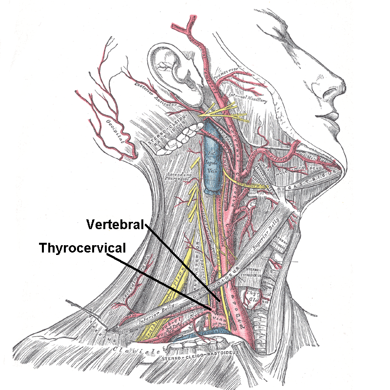
It gives off following important branches:
1. Vertebral artery
2. Thyrocervical trunk
Relations:
Anteriorly:
Moving medially to laterally are the common carotid arteries, vagus nerve and the internal jugular vein along with the cardiac branches of the vagus nerve and sympathetic nerves.
Posteriorly:
The apex of the lung and the dome of cervical pleura are located posteriorly. Also on the right side is the right recurrent laryngeal nerve.
Grey’s Anatomy 20th edition
Branches:
1. Vertebral artery:
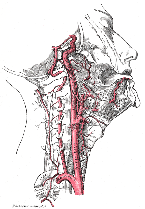
The vertebral artery arises from the upper margin of the first part of subclavian artery. It moves upwards in the neck, between the longus colli and the scalenus anterior muscles. On passing in front of the transverse process of the seventh cervical vertebra, it enters the foramina present in the transverse process of sixth cervical vertebra. It now passes upwards through the foramina in transverse processes of upper six cervical vertebra. It curves backwards and behind the lateral mass of the atlus, on emerging from the transverse process of the atlus. It now pierces the dura mater by passing medially to enter the vertebral canal. Ascending through the foramen magnum, enters the skull and supplies the brain.
The vertebral artery of both sides join together to form the basilar artery.
Relations:
Anteriorly:
Common carotid artery, thoracic duct crosses on the left side
Posteriorly:
Transverse process of seventh cervical vertebra, stellate ganglion, anterior rami of seventh and eighth cervical nerve.
The vertebral artery lies in front of the anterior rami of cervical nerves as it moves upwards through the foramina in the transverse processes.
Branches of Vertebral artery:
a. Spinal branches:
Spinal branches pass into the vertebral canal through the intervertebral foramina.
b. Muscular branches
Grey’s Anatomy 20th edition
2. Thyrocervical Trunk
Arising at the medial border of the scalenus anterior muscle, the thyrocervical trunk is a short trunk originating from the front of the first part of subclavian artery.
Branches of Thyrocervical Trunk:
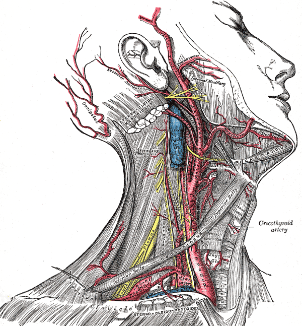
a. Inferior Thyroid Artery:
The inferior thyroid artery ascends to the level of the cricoid cartilage, along the medial border of the scalenus anterior muscle. Now it moves downwards after turning medially and moves behind the carotid sheath. The artery is related to the recurrent laryngeal nerve and supplies the thyroid on reaching the posterior border.
b. Superficial Cervical Artery
It enters the posterior triangle of the neck passing laterally across the scalenus anterior muscle. It is present in the lower part of the posterior triangle and disappears beneath the trapezius muscle.
c. Suprascapular Artery
It enters the posterior triangle of the neck passing laterally across the scalenus anterior muscle. It follows the suprascapular nerve to enter the supraspinous fossa taking part in the arterial anastomosis around scapula.
3. Internal Thoracic Artery
Arising from the lower border of the first part of subclavian artery, the internal thoracic artery descends in front of the pleura and behind the first costal cartilage in the thorax. The phrenic nerve crosses it obliquely from lateral to medial side. It ends by dividing into:
Second Part of Subclavian Artery
The part behind the scalenus anterior muscle is known as the second part of subclavian artery.
It gives off the costovervical trunk.
Costocervical Trunk
The costocervical trunk gives off:
a. Superior Intercostal Artery:
The posterior intercostal arteries of the first and second intercostal spaces are given off from the superior intercostal artery.
b. Deep Cervical Artery
The deep cervical artery gives off branches to the muscles of the back of the neck.
Relations:
Anteriorly:
Scalenus anterior muscle
Posteriorly:
Apex of lung and dome of cervical pleura
Third Part of Subclavian Artery:
Extending from the lateral border of the scalenus anterior muscle to the outer border of the first rib is the third part of subclavian artery. At the outer border of the first rib, the subclavian artery becomes the axillary artery.
The third part of subclavian artery carries the axillary sheath along with the brachial plexus of nerves. It enters the posteroinferior angle of the posterior triangle of neck. It disappears behind the middle of the clavicle.
Normally no branch arises from the third part of subclavian artery but occasionally the superficial cervical and the suprascapular artery arise from here.
 howMed Know Yourself


