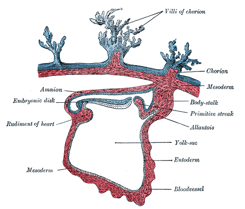
1) The amnio-ectodermal junction is the primitive umbilical ring.
2) Following structures pass through the ring at the 5th week of development:
a. Connecting stalk, containing Allantois and the umbilical vessels
b. Yolk stalk (vitelline duct), accompanied by vitelline vessels
c. Canal connecting the intraembryonic and extraembryonic cavities
3) The embryonic cavity enlarges rapidly and the amnion envelops the connecting and yolk sac stalks crowding them and give rise to primitive umbilical cord.
4) At the end of 3rd month loops with draw, cavity in the cord, chorionic cavity and yolk sac are obliterated.
5) When the allantois and the vitelline duct and its vessels are obliterated, all that remains in the cord are the umbilical vessels surrounded by the jelly of Wharton.
T.S OF UMBILICAL CORD
2cm in dia;50-60cm long
n Battledore placenta: Marginal insertion
n Velamentous insertion of cord: attached to fetal membranes
AMNIOTIC FLUID
n Amniotic cavity is filled with clear watery fluid
n Derived from maternal blood and amniotic cells
n 800-1000ml at 37 weeks
n By fifth month fetus swallows own amniotic fluid about 400ml a day
n Fetal urine is added daily in amniotic fluid in 5th month
Functions of Amniotic Fluid
– Absorb jolts
– Prevent adherence to the amnion
– Allows free fetal movements
– Helps in dilatation of cervical canal at the time of birth
Clinical correlates
1) Polyhydroamnios (more than1500-2000 ml)
n Causes: Idiopathic, maternal diabetes, congenital malformations, anencephaly, esophageal atresias.
2) Oligohydroamnios (less than 400 ml)
n Causes: Renal agenesis, premature rupture of membranes.
 howMed Know Yourself


