| Contents |
| 3rd Month |
| 4th and 5th month |
| 6th and 7th month |
| 9th month |
| Intrauterine Growth Restriction |
The transformation of embryo to fetus is gradual. The crown rump length or crown heel length is measured for finding out the age of the baby. Growth in length is quite evident during the 3rd, 4th and 5th months. Whereas, the increase in weight is most observed during the last 2 months of gestation.
Monthly Changes
The growth of the head during fetal period is slow compared to the rest of body. At the beginning of 3rd month, the head constitutes approximately ½ of crown – rump length (CRL). By the beginning of 5th month, the size of the head is about 1/3rd of crown – heel length (CHL). At birth is approximately 1/4th of the crown heel length..
Third Month (9-12 wks)
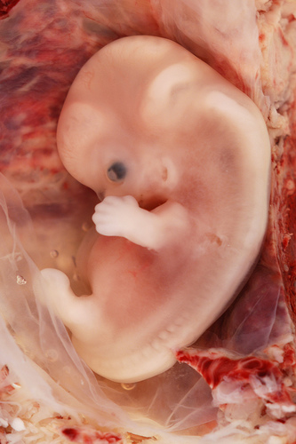
Photo by euthman. 9th week of development
Changes are observed in:
1. Face:
Face becomes more human looking and broad.
2. Head
Head is disproportionately large and makes up nearly half of the fetus’ size.
3. Eyes
Eyes are directed ventrally. The eyelids are fused.
4. Ears
Ears rise to their definitive position on the side of head
5. Limbs
Limbs reach their relative length
6. Primary Ossification Centers
Primary ossification centers are present in the long bones and skull by the 12th week.
7. Preumblical Herniation
During the 6th week, the intestinal loops cause a large swelling (physiological herniation) in the umbilical cord. By the 12th week, loops withdraw into the abdominal cavity.
8. Erythropoiesis
At 9th weeks, the liver is the major site for erythropoiesis. After 12th week, the activity is reduced in liver and begins in spleen.
9. External Genitalia
External genitalia appear to be well differentiated.
10. Muscular yActivity
Muscular activity can be evoked in aborted fetuses.
4th And 5th Months
Following changes are observed during the fourth and fifth weeks:
1. Length
The fetus rapidly grows in length and at the end of first half of intrauterine life, its crown rump length is approximately 150 mms, about ½ the total length of newborn.
2. Weight
The weight remains less than 0.5 kg.
3. Hair
Fine downy hairs (known as lanugo hair ) can be seen. Head and eye brow hairs become visible.
4. Movements
Fetal movements can be observed (which are quickening with time)
Mother may feel a fluttering in the lower abdomen
5. Skin
17 week fetus has thin skin with little connective tissue.
6th And 7th Months
1. Weight
During the 2nd half of intrauterine life, weight of the fetus increases. During last two and a half months 50% of the full term weight (approximately 3.2 kgs) is added.
2. Skin
During the 6th month, the skin of the fetus appears wrinkled and has reddish colour due to the absence of underlying connective tissue.
3. Respiratory and Nervous Systems
The respiratory and the central nervous system have not differentiated completely and coordination between the two systems is not yet well developed.
4. Length
By six and a half to seven months, fetus has developed a length of about 250 mm and has a weight of approximately 1.1 kg. The infant has a 90% chance of surviving, if it is born at this time.
5. Movements
Respiratory movements and sucking movements can be observed around this period of time.
6. Hearing
Some sounds can be heard by the fetus as well.
Ninth Month (33-36 Wks)
Skin
During ninth month the dull redness of the skin fades and the wrinkles smooth out. The lanugo hair begin to disappear
Body And Limbs
The body become rounded and the body fat increases
Vernix Caseosa
By the end of intrauterine life, the skin is covered by a whitish fatty substance
Skull
Skull has the largest circumference of all parts of the body, at the end of ninth month.
At the time of birth:
- The weight of a normal fetus is around 3 to 3.4 kgs
- Crown rump length is about 360 mms
- Crown heel length is about 500 mms.
Testes are in the scrotum in the males at the end of 9th month.
Time Of Birth
The date of birth can be indicated as 266 days, or 38 weeks, after fertilization. The sperm present in the reproductive tract up to 6 days prior to ovulation can survive to fertilize oocyte. The oocyte is usually fertilized within 12 hours of ovulation. This is why most pregnancies occur when sexual intercourse occurs within a 6-day period ending on the day of ovulation. The date of birth is calculated as 280 days or 40 weeks from the first day of last menstrual period. This method is fairly accurate as most fetuses are born within 10 to 14 days of the calculated delivery date. The babies are called premature, if they are born much earlier. If born later, they are called postmature.
Measurements And Characteristics Of Fetuses
Various measurements and external characteristics are useful for estimating the fetal age. Until the end of first trimester, crown rump length is the method of choice. In the second and third trimesters, several structures can be identified and measured ultrasonographically. Gestational sac appear on transabdominal sonography at 5th week (embryo at 7th week) and approx a week earlier by transvaginal ultrasound (TVS) (embryo at 6th week)
Ultrasonography Measurements
Ultrasonography measurements include:
- Biparietal diameter (BPD)
- Head circumference
- Abdominal circumference
- Femur length
- Foot length
- Fetal weight
Factors Influencing Fetal Growth
Following factors stimulate fetal growth:
- Glucose
- Amino acids
- Insulin (secreted by fetal pancreas)
- Insulin like growth factors
- Human growth hormone
Intrauterine Growth Restriction:
Intrauterine growth restriction or IUGR is a term applied to infants who are at or below the 10th percentile for their expected birth weight at a given gestational age. They are small for dates or small for gestational age. The incidence is 1 in 10 babies
Factors Causing IUGR:
Different factors causing intrauterine growth restriction include:
- Cigarette smoking
- Alcohol consumption
- Maternal malnutrition
- Faulty food habits
- Multiple pregnancies
- Drug addiction
- Impaired uteroplacental and fetoplacental blood flow (due to small chorionic vessels, severe hypotension, placental dysfunction or defect and renal diseases)
- Genetic factors and growth retardation (such as recessive genes, structural and numerical chromosomal aberrations)
 howMed Know Yourself


IUGR Ąπϑ SGA(small for gestationaal age are not the same
It is just mentioned that SGA may be associated with IUGR, which is known as ‘SGA associated with IUGR’. It’s not mentioned clearly, thanks for pointing it out.