The main arterial trunk which supplies oxygenated blood from the left ventricle of the heart to all tissues of the body is known as the aorta. Due to its extensiveness, it is divided into following parts, for the sole purpose of description:
1. Ascending aorta
2. Arch of aorta
3. Descending aorta
4. Abdominal aorta
Grey’s Anatomy 20th edition
1. Ascending aorta
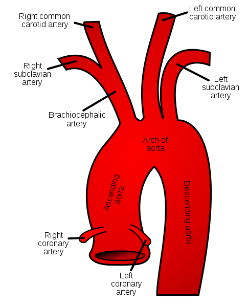
The ascending aorta arises from the base of the left ventricle. At the level of the sternal angle, it comes to lie behind the right half of sternum by moving upwards and forward. Here it becomes continuous with the arch of aorta.
The ascending aorta is surrounded by the fibrous pericardium and is enclosed by the sheath of pericardium along with the pulmonary trunk. The sinuses of aorta, which are the bulges located at the root, are located one behind each aortic valve cusp.
Photo by ZooFari
Branches:
1. Right coronary artery:
The right coronary artery arises from the anterior aortic sinus and supplies the right atrium, the right ventricle, parts of left atrium, left ventricle and the atrioventricular septum.
2. Left coronary artery
The left coronary artery arises from the left posterior aortic sinus and supplies the greater part of the left atrium, left ventricle and the ventricular septum.
Grey’s Anatomy 20th edition
2. Arch of aorta
The ascending aorta leads into the arch of aorta. The arch of aorta lies behind the manubrium sterni. It arches upwards, backwards and to the left, lying in front of the trachea. It continues downwards and to the left of trachea. It becomes continuous with the descending aorta, at the level of the sternal angle.
Branches:
1. Brachiocephalic artery:
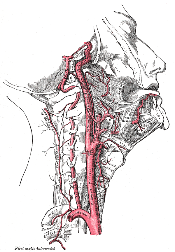
The convex surface of the arch of aorta gives rise to the brachiocephalic artery. It continues upwards and to the right side of the trachea, dividing into two terminal branches behind the right sternoclavicular joint:
b. Right common carotid artery
2. Left common carotid artery:
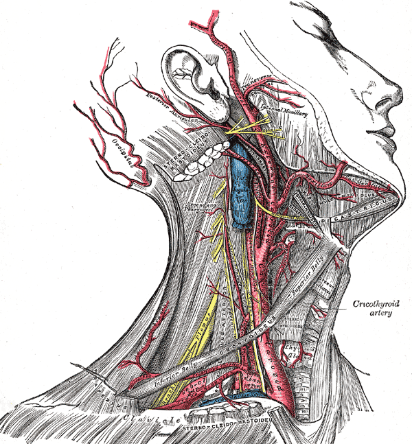
The left common carotid artery arises on the left side of the brachiocephalic artery from the convex surface of the arch of aorta. It enters the neck behind the left sternoclavicular joint by moving upwards and to the left of trachea.
3. Left subclavian artery:
The left subclavian artery arises behind the left common carotid artery from the arch of aorta. It enters the root of the neck by moving upwards along the left side of the trachea. It arches over the apex of the left lung.
3. Descending Thoracic Aorta:
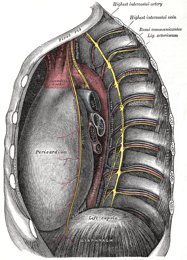
Located in the posterior mediastinum, the descending thoracic aorta is a continuation of the arch of aorta, beginning on the left side of the lower border of the body of fourth thoracic vertebra, which is located opposite the sternal angle.
The descending thoracic aorta runs downwards, moving forward and medially and reaching the anterior surface of the vertebral comumn. It continues on to the abdominal aorta by passing behind the diaphragm, at the level of the twelfth thoracic vertebra. The aorta passes through the aortic opening.
Grey’s Anatomy 20th edition
Branches:
1. Posterior intercostal arteries:
The lower nine intercostal spaces are provided with the posterior intercostal arteries on each side.
2. Subcostal arteries:
The subcostal arteries enter the abdominal wall by running along the lower border of the twelfth rib.
3. Pericardial branches
4. Esophageal branches
5. Bronchial arteries
4. Abdominal Aorta:
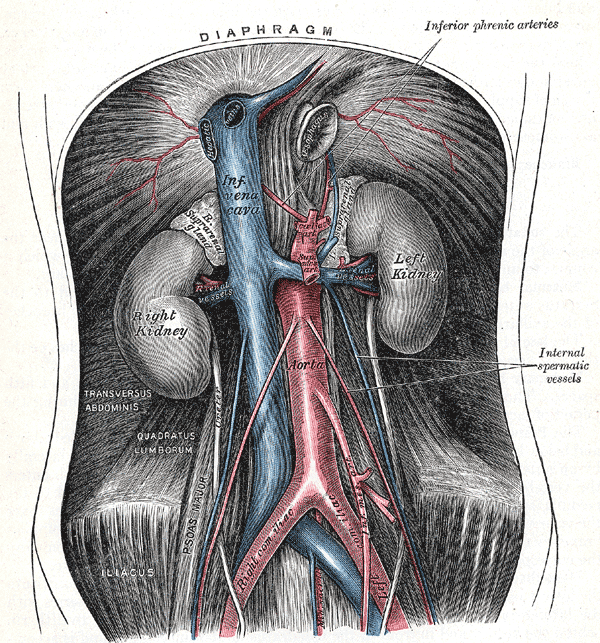
The abdominal aorta is the continuation of the descending thoracic aorta beginning at the aortic opening in the diaphragm, at the level of the twelfth thoracic vertebra. It descends on the anterior surface of the bodies of lumbar vertebra behind the peritoneum.
It divides into two common iliac arteries at the level of the fourth lumbar vertebra.
Grey’s Anatomy 20th edition
Relations:
On the right lie the inferior vena cava, the beginning of azygos vein and the cisterna chyli.
On the left lies the left sympathetic trunk.
Branches:
1. Anterior visceral branches:
a. Celiac artery
b. Superior mesenteric artery
c. Inferior mesenteric artery
2. Lateral visceral branches
a. Suprarenal artery
b. Renal artery
c. Testicular or ovarian artery
3. Lateral abdominal wall branches:
a. Inferior phrenic artery
b. Four lumbar arteries
4. Terminal branches:
b. Median sacral artery:
The median sacral artery arises at the bifurcation of the aorta. It moves downwards on the anterior surface of the sacrum and the coccyx.
 howMed Know Yourself



really helpful….good work
really good helpful, can improve still