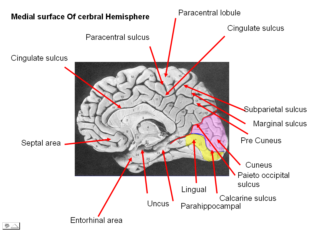
• The cerebrum is the largest part of the brain, situated in the anterior and middle cranial fossae of the skull and occupying the whole concavity of the vault of the skull.
• It may be divided into two parts:
– the diencephalon, which forms the central core, and
– the telencephalon, which forms the cerebral hemispheres.
Diencephalon
• The diencephalon consists of the third ventricle and the structures that form its boundaries
• The diencephalon can be divided into four major parts:
• the thalamus,
• the subthalamus,
• the epithalamus, and
• the hypothalamus.
Telencephalon
• The cerebral hemispheres are the largest part of the brain
• They are separated by a deep midline sagittal fissure, the longitudinal cerebral fissure
• In the depths of the fissure, the great commissure, the corpus callosum, connects the hemispheres across the midline
• A second horizontal fold of dura mater separates the cerebral hemispheres from the cerebellum and is called the tentorium cerebelli.
CEREBRAL WHITE MATTER
Structure
• White matter is composed of bundles of myelinated nerve cell processes (or axons), which connect various grey matter areas (the locations of nerve cell bodies) of the brain to each other, and carry nerve impulses between neurons.
• Cerebral– and spinal white matter do not contain dendrites, which can only be found in grey matter along with neural cell bodies, and shorter axons.
• The white matter is white because of the fatty substance (myelin) that surrounds the nerve fibers (axons).
• This myelin is found in almost all long nerve fibers, and acts as an electrical insulation.
• This is important because it allows the messages to pass quickly from place to place
Functions
• The white matter is the tissue through which messages pass between different areas of gray matter within the nervous system.
• Using a computer network as an analogy, the gray matter can be thought of as the actual computers themselves, whereas the white matter represents the network cables connecting the computers together.
Types of White Fibers
• Commissural Fibers
• Association Fibers
• Projection Fibers
Commissural Fibers
• Commissure fibers essentially connect corresponding regions of the two hemispheres.
– Corpus callosum
– Anterior Commissure
– Posterior Commissure
– Fornix
– Habenular Commissure
• The corpus callosum, the largest commissure of the brain, connects the two cerebral hemispheres.
• It lies at the bottom of the longitudinal fissure.
For purposes of description, it is divided into
• the rostrum,
• the genu,
• the body, and
• the splenium.
Extensions
• Forceps minor
• Radiation of corpus callosum
• Forceps major
• Tapetum
The Anterior Commissure
• It is a bundle of nerve fibers connecting the two cerebral hemispheres across the midline, and placed in front of the columns of the fornix
• It crosses the midline in the lamina terminalis
Connections
• When traced laterally, a smaller or anterior bundle curves forward on each side toward the anterior perforated substance and the olfactory tract.
• A larger bundle curves posteriorly on each side and grooves the inferior surface of the lentiform nucleus to reach the temporal lobes.
The Posterior Commissure (Epithalamic Commisure)
• Is a rounded band of white fibers crossing the midline on the dorsal aspect of the upper end of the cerebral aqueduct.
• It is related to the inferior part of the stalk of the pineal gland.
Origin & Functions
• Most of them have their origin in a nucleus, the nucleus of the posterior commissure (nucleus of Darkschewitsch), which lies in the central gray substance of the upper end of the cerebral aqueduct
• Some fibers are probably derived from the posterior part of the thalamus and from the superior colliculus, whereas others are believed to be continued downward into the medial longitudinal fasciculus.
• The posterior commissure interconnects the pretectal nuclei, mediating the consensual pupillary light reflex.
The Fornix
• The fornix (Latin, “vault” or “arch”) is a C-shaped bundle of fibres (axons) in the brain, and carries signals from the hippocampus to the mammillary bodies.
• The white matter that covers the superior surface of hippocampus is called Alveus.
• These fibers begin in the hippocampus & converge on its medial border to form a bundle called Fimbria;
• these fibers leave the posterior end of hippocampus & separate on left and right side & are called the crus of the fornix (Posterior column of Fornix).
• The bundles of fibers come together in the midline of the brain, forming the body of the fornix.
• The inferior edge of the septum pellucidum (a membrane that separates the two lateral ventricles) is attached to the upper face of the fornix body.
• As the two crura come together, they are connected by transverse fibers called Commissure of Fornix which decussate & join the hippocampi of two sides
• The body of the fornix travels anteriorly and divides again near the anterior commissure.
• The left and right parts reseparate and form two anterior columns of Fornix.
• The fibres of each side continue through the hypothalamus to the mammillary bodies; then to the anterior nuclei of thalamus,
The habenular commissure
• Is situated in front of the pineal gland that connects the habenular nucleus on one side of the diencephalon with that on the other side.
• It crosses the mid line in the superior part of root of pineal stalk.
• Receive afferents from hippocampus & amygdaloid nuclei which run in stria medullaris thalami.
Association Fibers
Association fibers are nerve fibers that essentially connect various cortical regions within the same hemisphere
• Short Association fibers
• Long Association fibers
The long association fibers
• The uncinate fasciculus connects the first motor speech area and the gyri on the inferior surface of the frontal lobe with the cortex of the pole of the temporal lobe.
• The cingulum is a long, curved fasciculus lying within the white matter of the cingulate gyrus .It connects the frontal and parietal lobes with parahippocampal and adjacent temporal cortical regions.
• The superior longitudinal fasciculus is the largest bundle of nerve fibers. It connects the anterior part of the frontal lobe to the occipital and temporal lobes.
• The inferior longitudinal fasciculus runs anteriorly from the occipital lobe, passing lateral to the optic radiation, and is distributed to the temporal lobe.
• The fronto-occipital fasciculus connects the frontal lobe to the occipital and temporal lobes. It is situated deep within the cerebral hemisphere and is related to the lateral border of the caudate nucleus.
Projection Fibers
• Afferent and efferent nerve fibers passing to and from the brainstem to the entire cerebral cortex.
• Pass between large nuclear masses of gray matter within the cerebral hemisphere.
• At the upper part of the brainstem, these fibers form a compact band known as the Internal Capsule
• Once the nerve fibers have emerged superiorly from between the nuclear masses, they radiate in all directions to the cerebral cortex. These radiating projection fibers are known as the Corona Radiata
The internal capsule
• Is an area of white matter in the brain that separates the caudate nucleus and the thalamus from the lenticular nucleus. The internal capsule contains both ascending and descending axons.
• Is an area of white matter in the brain that separates the caudate nucleus and the thalamus from the lenticular nucleus. The internal capsule contains both ascending and descending axons.
• V-shaped when cut both coronally (on the same plane as the face) and horizontally/transversely
• When cut horizontally:
• the bend in the V is called the “genu“.
• the part in front of the genu is the “anterior limb“.
• the part behind the genu is called the “posterior limb”
• There is also a retrolenticular and a sublenticular part to the internal capsule
• The posterior limb of the internal capsule contains corticospinal fibers and sensory fibers from the body.
• The genu contains corticobulbar fibers, which run between the cortex and the brainstem.
• The anterior limb of the internal capsule contains:
• 1) frontopontine (corticofugal) fibers project from frontal cortex to pons;
• 2) thalamocortico fibers connect the medial and anterior nuclei of the thalamus to the frontal lobes (these are severed during a prefrontal lobotomy).
• The retrolenticular part contains fibers from the optic system, coming from the lateral geniculate nucleus of the thalamus. More posteriorly, this becomes the optic radiation.
• Some fibers from the medial geniculate nucleus (which carry auditory information) pass mostly in the sublenticular part.
CLINICAL ANATOMY
• Commissures of the Cerebrum
The major commissure is the large corpus callosum. The majority of the fibers within the corpus callosum interconnect symmetrical areas of the cerebral cortex. Because it transfers information from one hemisphere to another, the corpus callosum is essential for learned discrimination, sensory experience, and memory.
• Occasionally, the corpus callosum fails to develop, and in these individuals, no definite signs or symptoms appear.
• Should the corpus callosum be destroyed by disease in later life, however, each hemisphere becomes isolated, and the patient responds as if he or she has two separate brains. The patient’s general intelligence and behavior appear normal, since over the years both hemispheres have been trained to respond to different situations.
• If a pencil is placed in the patient’s right hand (with the eyes closed), he or she will recognize the object by touch and be able to describe it. If the pencil is placed in the left hand, the tactile information will pass to the right postcentral gyrus. This information will not be able to travel through the corpus callosum to the speech area in the left hemisphere; therefore, the patient will be unable to describe the object in his or her left hand.
Lesions of the Internal Capsule
• Arterial Hemorrhage caused by Atheromatous Degeneration in an artery in a patient with high blood pressure.
• Because of the high concentration of important nerve fibers within the internal capsule, even a small hemorrhage can cause widespread effects on the contralateral side of the body.
• Not only is the immediate neural tissue destroyed by the blood, which later clots, but also neighboring nerve fibers may be compressed or be edematous.
Alzheimer Disease
• Early memory loss, a disintegration of personality, complete disorientation, deterioration in speech, and restlessness are common signs. In the late stages, the patient may become mute, incontinent, and bedridden and usually dies of some other disease.
• so-called senile plaques are found in the atrophic cortex.
• In the center of each plaque is an extracellular collection of degenerating nervous tissue, with an excess of intracellular neurofibrils that are tangled and twisted, forming neurofibrillary tangles.
Histological slides are available here.
 howMed Know Yourself


Pretty nice post. I just stumbled upon your blog and wanted to say that I have really enjoyed browsing your blog posts. In any case I’ll be subscribing to your feed and I hope you write again soon!