Principle
Most bacteria can be differentiated by the gram reactions due to difference in their cell wall structure.
Gram positive bacteria
Cell wall has a large amount of peptidoglycan.
Gram negative bacteria
They have less amount of peptidoglycan in their cell wall. They have lipopolysaccharide containing a compound known as lipid A or endotoxin.
The organisms that retain purple color with crystal violet and are not decolorized by acetone iodine are called gram positive bacteria.
The organisms that loose their color of crystal violet after being treated with acetone iodine are called gram negative bacteria.
Procedure
- Fix the smear of specimen (sputum, fluid or culture) either by heating or alcohol fixation.
- Cover the fixed smear with crystal violet stain for 30 seconds and then wash off with water.
- Cover the smear with lugol iodine for 30 seconds and wash it off with water.
- Decolorize by pouring acetone iodine (5 seconds) and rapidly wash off.
- Now cover the slide with carbol fuschin for 30 seconds and wash it off with water.
- Allow the slide to air dry, observe under oil immersion lens.
- The report should include the following:
- Number of bacteria present, whether many, moderate, few or scanty.
- Whether gram positive or gram negative
- Morphology of bacteria, whether cocci, bacilli and their arrangement.
- Presence of pus cells
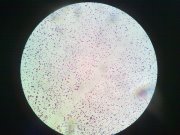
Gram Positive Cocci
False Gram Negative Staining
- Cell wall damaged due to antibiotic therapy or excessive heat fixation
- Over decolorization with acetone iodine
- Use of old iodine which is a poor mordant.
 howMed Know Yourself

Thanks alot for this piece of info.
It made me to understand it better.
thanks for contribution
Thanks for your contribution, I really appreciate. I found this article very helpul.
Thx a lot to help me to undrstood gram’s bacterias.
Short nd easy way to understand the bacterial staining procedure….gud work