Clinical Significance
1. Urine testing provides rapid, valuable and reliable information into the health status of the patient.
2. Urine provides a valuable index to many normal or pathological mechanisms. Urine is composed of various organic and inorganic substances produced in the body and are required to be excreted.
3. These substances are produced as a result of either normal or abnormal body metabolism.
Types of Urine Specimens:
1. First morning specimen:
It provides more concentrated specimen and is appropriate for nitrite and protein estimation and good for microscopic examination.
2. Random specimen:
It is the most common and convenient sample. It is good for observing physical characteristic, chemical analysis and identification of casts, crystals and cells
3. Second voided specimen:
It provides good correlation of urine glucose with blood glucose
4. Post-prandial:
It is good for glucose and protein estimation
5. Timed specimen:
It is combination of all voiding over a length of time.
Two hour specimen is good for urobilinogen and
Twenty four hour urine sample is good for quantitative urinary components estimation
Physical Examination:
1. Volume: Measure the exact volume of urine and report the same if requested.
2. Odour: Smell the urine immediately after opening the container and report the characteristic smell.
3. Color: Observe the color of the urine and report the same
4. Appearance: Observe the appearance of urine and report. The appearance ranges from clear when freshly voided and becomes cloudy on standing
5. pH: Dip the Uristix reagent strip and remove the excess of urine by touching the strip with the edges of container. Compare the color and supplied color chart and record pH.
Urinary pH can be checked with indicator paper strip. Strips used carry indicators methyl red (red strips) and bromothymol blue (blue strip), which provide a pH range of 5.0 – 9.0.
Normal pH is acidic (5.0 – 6.5)
6. Specific gravity: Dip the Uristix reagent strip and remove the excess of urine by touching the strip with the edges of container. Compare the colour and supplied colour chart and record specific gravity.
Urine specific gravity can also be measured by either Urinometer or Refrectometer.
Chemical Examination:
1. Proteins: Dip the Uristix reagent strip and remove the excess of urine by touching the strip with the edges of container. Compare the colour and supplied colour chart and record the semi quantitative result of protein estimation.
Heat and turbidimetric methods can also be done to quantitate the proteins in the urine. Turbidimetric method can be done by heating (boiling), heat and acetic acid, sulfosalicylic acid test and nitric ring test.
2. Glucose: Dip the Uristix reagent strip and remove the excess of urine by touching the strip with the edges of container. Compare the colour and supplied colour chart and record the amount of glucose.
Non-specific reduction method like Benedict’s test can also be used for screening of urine for reducing substances.
Microscopy:
Microscopy is and essential component of urinalysis. Following examination procedures are required:
Direct examination:
1. Light Microscopy: Using light microscopy observe for various parasites, ova or larvae of parasites, RBCs, leukocytes, casts, epithelial cell and crystals.
2. Dark ground illumination: This is required when organisms like Leptospira are expected.
Staining or urine sediment
a. Methylene blue
b. Sternheimer-Malbin stain
c. Stains for fat cells:
Sudan III
Sudan IV
Oil red O
d. Acid Fast Stain
ZN stain
Kanyoun stain
- Mix the specimen well.
- Take 10 – 15ml of urine specimen in centrifuge tube
- Centrifuge at 1000 rpm for 3 minutes
- Invert the tube to pour off the supernatant
- Mix the sediment with small amount of urine left over in the tube by holding the top of the tube with one hand and striking the bottom with the finger of the other.
- Pour a drop on a clean glass slide and cover with a cover slip
- Examine under subdued light, first scanning whole area with low power (10X) objective and then with high power objective (40X)
- Amorphous deposits can be removed by adding a small drop of 10% acetic acid if present in excess
Observe for the presence of following structures and report:
- Leuckocytes
- Erythrocytes
- Casts
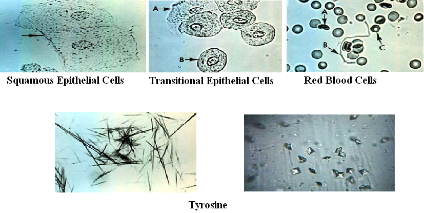
- Epithelial cells
- Crystals
- Amorphous deposits
- Squamous epithelial cells are large (40 – 60 µm), flat, irregularly-shaped cells with abundant cytoplasm and a small round nucleus
- Transitional epithelial cells are a little smaller than squamous epithelial cells ( 20 – 40 µm).
- The center pallor of most of the RBCs is quite evident.
- Tyrosine appears as fine, delicate needles in clusters or sheaves
Casts:
As the urine in the tubules becomes concentrated, as urine stasis occurs or when the pH is very low, the protein forms fibrils that attach to the lumen cells. The fibrils intertwine to eventually form the cast itself which is washed out into the urine as flow increases.
Want a clearer concept, also see
Lecture on Urine Routine Examination
Article on Urine Routine Examination
 howMed Know Yourself


