The brain is one of the most metabolically active organs in the body, receiving 17% of the total cardiac output and about 20% of the oxygen available in the body.
The brain receives it’s blood from
two pairs of arteries, the carotid and vertebral. About 80% of the brain’s
blood supply comes from the carotid, and the remaining 20% from the vertebral.
THE VERTEBROBASILAR SYSTEM
The vertebral arteries originate from the subclavian artery, and ascend through the transverse foramen of the upper six cervical vertebra. At the upper margin of the Axis (C2) it moves outward and upward to the transverse foramen of the Atlas (C1). It then moves backwards along the articular process of atlas into a deep groove, passes beneath the atlanto-occipital ligament and enters the foramen magnum. The arteries then run forward and unite at the caudal border of the pons to form the basilar artery.
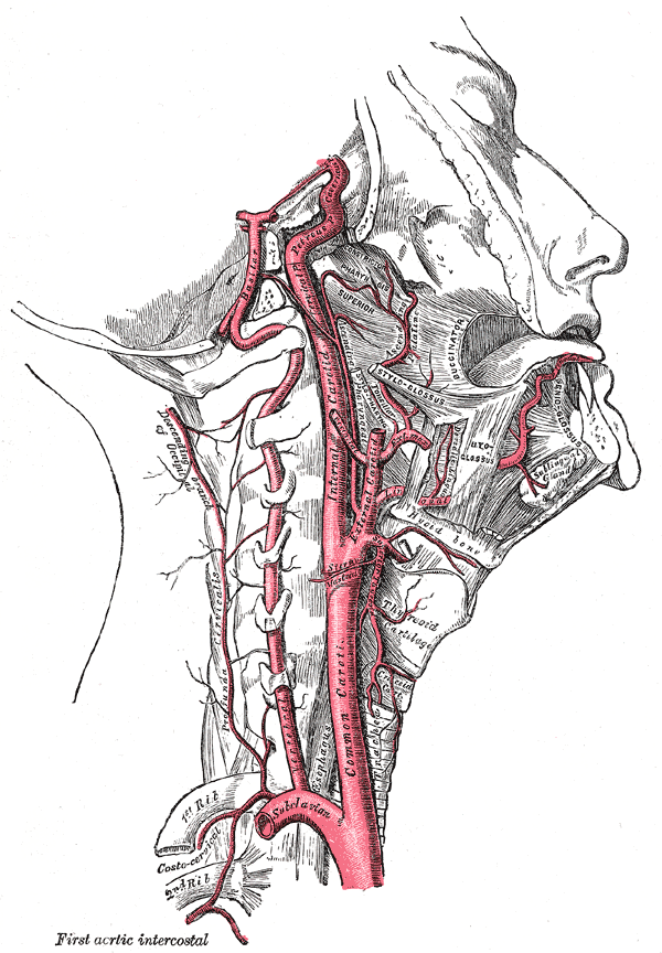
INTERNAL CAROTID ARTERY
• Begins at bifurcation of CCA
• Ascends the neck
• Perforates the base of skull & enters carotid canal
• Passes horizontally forward through cavernous sinus
• Emerges on medial side of anterior clinoid process by perforating the dura mater
• Enters the subaracnoid space & turns posteriorly
• Reaches medial end of lateral cerebral sulcus
BRANCHES OF ICA
• Ophthalmic
• Posterior communicating
• Anterior choroidal
• Anterior cerebral
• Middle cerebral
OPTHALMIC ARTERY
• Enters the orbit through optic canal
• Area of distribution:
• Eye & other orbital structures
• Frontal area of scalp
• Ethmoid & Frontal air sinuses
• Dorsum of nose
POSTERIOR COMMUNICATING ARTERY
• Originate close to the terminal bifurcation of ICA
• Runs posteriorly & join circle of Willis
Anterior Choroidal
• Arises directly from ICA
• Runs backwards in relation to optic tract
• Enters the inferior horn of lateral ventricle through choroid fissure
• Ends in the choroid plexus
• Area of distribution:
• Crus cerebri, LGB, Optic tracts & Internal capsule
Anterior cerebral
• Smaller terminal branch of ICA
• Runs forward & medially superior to the optic nerve & enters the longitudinal fissure of cerebrum
• Joined by ACA of opposite side by Anterior communicating artery
• It curves backward over the corpus callosum & anastomose with PCA
Area Supplied By Anterior cerebral
Cortical branches:
• All the medial surface of cerebral cortex upto parietooccipital sulcus
• Strip of cortex about 1 inch wide on adjoining superolateral surface
• Supplies leg area of precentral gyrus
• Olfactory bulb & tract on inferior surface
Central branches:
• Pierces anterior perforated substance & supply parts of lentiform & caudate nuclei & internal capsule
• The recurrent artery of Heubner irrigate the internal capsule
Occlusion of ACA
• Occlusion of the anterior cerebral artery may result in the following defects
• Paralysis of the contralateral foot and leg
• Sensory loss in the contralateral foot and leg
MIDDLE CEREBRAL ARTERY
• Largest branch of ICA, runs laterally in the lateral sulcus
Areas supplied by the middle cerebral artery include:
Cortical branches:
• Entire lateral surface of hemisphere except for the narrow strip of cortex supplied by ACA; occipital pole & inferior part of temporal lobe supplied by PCA
Central branches:
• Enter the anterior perforated substance & supply part of internal capsule & basal ganglia
Occlusion of the middle cerebral artery
• Occlusion of the middle cerebral artery may result in the following defects:
• Paralysis of the contralateral face and limbs.
• Sensory loss in the contralateral face and limbs.
• Aphasia e.g. Broca’s, Wernicke’s when the dominant hemisphere (usually the left hemisphere for right handed individuals) is affected
Circle of Willis
• Lies in the interpeduncular fossa at the base of brain
• Anastomosis b/w two ICA’s & two Vertebral arteries
• Allows blood to be distributed to any part of both hemispheres
• Cortical & central branches arise from the circle & supply brain substance
• The Circle of Willis, long considered to be an important anatomic vascular formation, provides backup circulation to the brain. In case one of the supply arteries is occluded, the Circle of Willis provides interconnections between the internal carotid arteries and basilar artery along the floor of the cerebral vault, providing blood to tissues that would otherwise become ischemic.
• Normally, there is little or no blood flow around this circle.
• However, if one of the major vessels becomes occluded within the circle or proximal to it, the communicating arteries may allow anastomotic flow and prevent neurological damage.
• There are small arteries that arise from the arterial circle and from the major cerebral arteries.
• These central or penetrating arteries from 4 groups:
• The anteromedial;
• The anterolateral;
• The posteromedial;
• The posterolateral.
ANTEROMEDIAL GROUP
• Anteromedial Group
• These arteries supply:
• The septal area;
• And preoptic and anterior regions of the hypothalamus.
• This group also includes the medial striate artery (recurrent artery of Heubner) which arises from the anterior cerebral artery.
• It moves sharply backwards to supply more lateral regions:
• The head of caudate;
• And the anterior limb of the internal capsule.
ANTEROLATERAL GROUP
• Anterolateral Group
• This is also called the lateral striate or lenticulostriate arteries.
• They supplies:
• The head of caudate nucleus;
• The claustrum;
• The external capsule;
• The putamen;
• The lateral globus pallidus;
• And most of the internal capsule (except ventral part of the posterior limb which is supplied by the anterior choroidal artery).
Posteromedial Group
• These arteries supply:
• The medial parts of the midbrain;
• The anterior and medial thalamus;
• The subthalamic regions;
• And the middle and posterior hypothalamus.
POSTEROLATERAL GROUP
• These are also called the thalamogeniculate arteries.
• They supply:
• The lateral part of the midbrain;
• And the posterior aspect of the thalamus (including the geniculate bodies, VPL and pulvinar).
THE VEINS OF THE BRAIN
The veins of the brain possess no valves, and their walls, owing to the absence of muscular tissue, are extremely thin. They pierce the arachnoid membrane and the inner or meningeal layer of the dura mater, and open into the cranial venous sinuses.
Cerebral venous drainage
• The venous drainage of the cerebrum can be separated into two subdivisions: superficial and deep.
• The superficial system is composed of dural venous sinuses
• The deep venous drainage is primarily composed of traditional veins inside the deep structures of the brain, which join behind the midbrain to form the Vein of Galen. This vein merges with the Inferior sagittal sinus to form the Straight sinus which then joins the superficial venous system mentioned above at the Confluence of sinuses.
The cerebral veins
• Are divisible into
• External cerebral veins
• Internal cerebral veins
Accordingly as they drain the outer surfaces or the inner parts of the hemispheres.
• The external veins are the superior, inferior, and middle cerebral veins
EXTERNAL CEREBRAL VEINS
- The Superior Cerebral Veins, eight to twelve in number, drain the suprolateral and medial surfaces of the hemispheres. They open into the superior sagittal sinus
- The Superficial Middle Cerebral Vein / superficial Sylvian vein begins on the lateral surface of the hemisphere, and, running along the lateral cerebral fissure, ends in the cavernous or the sphenoparietal sinus. Through the superior & inferior anastomotic veins it communicates with the SSS & Transverse sinus
3.The Inferior Cerebral Veins of small size, drain the under surfaces of the hemispheres. Those on the orbital surface of the frontal lobe join the superior cerebral veins, and through these open into the superior sagittal sinus; those of the temporal lobe anastomose with the middle cerebral and basal veins, and join the cavernous, sphenoparietal, and superior petrosal sinuses.
- Deep middle cerebral vein drains the surface of insula & terminate in basal vein and runs in the lower part of the lateral cerebral fissure
- Anterior cerebral veins are small veins which drain the corpus callosum & anterior part of medial surface of hemisphere. They terminate in the basal vein
THE INTERNAL CEREBRAL VEINS
• Drains the deep parts of the hemisphere and are two in number; each is formed at the interventricular foramen by the union of the thalmostriate veins and choroid veins.
• They run backward parallel with one another, between the layers of the tela chorioidea of the third ventricle, and unite beneath the splenium of the corpus callosum, to form a short trunk, the great cerebral vein which ends in straight sinus, just before their union each receives the corresponding basal vein
TERMINAL VEINS
1)BASAL VEIN
• The basal vein is formed at the anterior perforated substance by the union of three veins
- A small anterior cerebral vein
- The deep middle cerebral
- The inferior striate veins, which leave the corpus striatum through the anterior perforated substance.
• The basal vein passes backward around the cerebral peduncle, and ends in the internal cerebral vein (vein of Galen)
• Its tributaries are from the interpeduncular fossa, the inferior horn of the lateral ventricle, the hippocampal gyrus, and the mid-brain
2)GREAT CEREBRAL VEIN OF GALEN
Formed by the union of the two internal cerebral veins, is a short median trunk which curves backward and upward around the splenium of the corpus callosum and ends in the anterior extremity of the straight sinus.