It is an areolar tissue – supports muscles, vessels n viscera of neck
4 parts
n Investing layer
n Pretracheal layer
n Prevertebral layer
n Carotid sheath
Investing Layer of Deep Cervical Fascia
n Completely encircles the neck like a collar
n Splits to enclose sternocleidomastoid & trapezius muscles and blends posteriorly with ligamentum nuchae which is attached to spines of cervical vertebrae.
n Splits to enclose submandibular and parotid glands
n Attached anteriorly to the hyoid bone
n Attached above to lower border of mandible, zygomatic arch, base of skull, mastoid process, superior nuchal line and external occipital protuberance
n Attached below to manubrium sterni, clavicle and acromion
n b/w angle of mandible & tip of mastoid process, inv layer is strong n splits to enclose Parotid;
n Superficial part extends superiorly as Parotidomasseteric fascia n reaches upto zyg arch
n Deep part extends to skull base; b/w styloid n angle of mandible, its thickened as Stylomandibular ligament.
its attached to both clavicles n to jugular (suprasternal) notch by 2 layers into which it splits a short distance above them. Layers are attached to ant n post borders of jugular notch, enclosing suprasternal space.
Contents of suprasternal space
n Anterior jugular veins (lower parts)
n Anastomotic arch between them
n Sternal heads of sternocleidomastoids
n A Lymph node (sometimes)
Pretracheal Layer of Deep Cervical Fascia
n Thin fascia, lying deep to infrahyoid strap muscles (sternothyroid, sternohyoid and omohyoid)
n It splits to enclose the thyroid gland, to which it is not adherent except b/w isthmus and 2nd,3rd & 4th tracheal rings
n It also encloses the para thyroid glands & the infrahyoid strap muscles
n Attached above to the thyroid and cricoid cartilage
n Below it extends into thorax & blends with adventitia of aortic arch and the fibrous pericardium
n Laterally it blends with carotid sheath and with the Investing layer of deep cervical fascia
Prevertebral Layer of Deep Cervical Fascia
n It covers the prevertebral muscles-namely, the longus capitis & longus cervicis.
n Passes around the neck to be attached to ligamentum nuchae
n In the posterior triangle, forms the fascial floor.
n All cervical nerve roots lie deep to it; so cervical plexus & trunks of brachial plexus lie deep to it.
n Lymph nodes of posterior triangle and accessory nerve lie superficial to it.
n Below it enters the thorax and blends with the anterior longitudinal ligament on the body of T4 vertebra.
n As subclavian A & brachial plexus emerge in the interval between scalenus anterior & medius, they carry sheath of fascia which extends into the axilla– called Axillary sheath
n The interval between the pharynx and the prevertebral fascia is called the retropharyngeal space
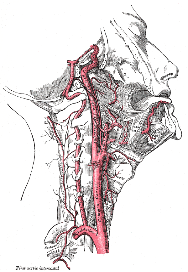
Carotid sheath
Attached to the base of skull at the margins of carotid canal & jugular fossa and is continued downwards along the vessels to blend with adventitia of aortic arch.
It is thin where it overlies IJV, allowing the vein to dilate during increased blood flow.
Contents are
n Common carotid artery
n Internal carotid artery
n Internal jugular vein
n Vagus nerve
Clinical Significance
n Important for surgeon
n Spread of infections in tissue spaces (visceral, retropharyngeal, submandibular & masticatory)- tough fascia can determine the direction of spread of infection. Pus from retrophar space to sup. mediastinum.
n Lower molar dental infections spread medially from mandible into submandibular & masticatory spaces and pushes tongue forward & upward.
n Further spread downward involve visceral space and lead to edema of vocal cords and air way obstruction.
n Ludwig’s angina -an acute, rare but severe form of infection of submandibular space, secondary to dental infection.
Chronic infection of fascial spaces of neck
n Tuberculous infection of deep cervical nodes- pus first limited by investing layer;later this becomes eroded at one point, pus comes to less restricted superficial fascia. Dumbbell or collar stud abscess is now present.
n Clinician should not forget deeply placed abscess


This is such a great resource that you are providing and you give it away for free. I enjoy seeing websites that understand the value of providing a prime resource for free. I truly loved reading your post. Thanks!
I agree with your thoughts here and I really love your blog! I’ve bookmarked it so that I can come back & read more in the future.