n The spinal cord is roughly cylindrical in shape.
n It begins superiorly at the foramen magnum in the skull.
n It terminates inferiorly in the adult at the level of the lower border of the L1.
n In the young child, it usually ends at the upper border of L3.
MENINGES
q It is surrounded by the dura mater, the arachnoid mater, and the pia mater.
q Further protection is by the CSF which surrounds the spinal cord .
ENLARGEMENTS
q Cervical & lumbar enlargements
q In the cervical region, it gives origin to the brachial plexus
q In the lower thoracic and lumbar regions, it gives origin to the lumbosacral plexus
CONUS MEDULLARIS:
Inferiorly, the spinal cord tapers off into the conus medullaris.
q FILUM TERMINALE:
From the apex of conus medullaris; a prolongation of the pia mater, descends to be attached to the posterior surface of coccyx.
FISSURE & SULCI
q In the midline anteriorly, the anterior median fissure.
q On the posterior surface, a shallow furrow, the posterior median sulcus.
SPINAL NERVES
q Along the entire length of the spinal cord are attached 31 pairs of spinal nerves
Cervical …… 8
Thoracic …… 12
Lumbar …… 5
Sacral …… 5
Coccygeal …..1
- Each connects to the spinal cord by 2 roots – dorsal and ventral.
- Each root forms from a series of rootlets that attach along the whole length of the spinal cord segment.
- Ventral roots are motor while dorsal roots are sensory.
The 2 roots join to form a spinal nerve prior to exiting the vertebral column.
Almost immediately after emerging from its intervertebral foramen, a spinal nerve will divide into a dorsal ramus, a ventral ramus, and a meningeal branch that re-enters and innervates the meninges and associated blood vessels.
- Each ramus is mixed.
- Joined to the base of the ventral rami of spinal nerves in the thoracic region are the rami communicantes. These are sympathetic fibers.
- Dorsal rami supply the posterior body trunk whereas the thicker ventral rami supply the rest of the body trunk and the limbs.
Plexuses
- Except for T2 to T12, all ventral rami branch extensively and join one another lateral to the vertebral column forming complicated nerve plexuses.
- Within a plexus, fibers from different rami criss cross each other and become redistributed.
Internal Structure:
The spinal cord is composed of an inner core of gray matter, which is surrounded by an outer covering of white matter.
GRAY MATTER
- On cross section; the gray matter is seen as an H-shaped pillar with anterior and posterior gray columns, or horns, united by a thin gray commissure containing the small central canal.
- A small lateral gray column or horn is present in the thoracic and upper lumbar segments of the cord.
- The amount of gray matter present at any given level of the spinal cord is related to the amount of muscle innervated at that level.
- Thus, its size is greatest within the cervical and lumbosacral enlargements of the cord.
WHITE MATTER
q The white matter, may be divided into anterior, lateral, and posterior white columns or funiculi.
q The anterior column on each side lies between the midline and the anterior nerve roots;
q The lateral column lies between the anterior and the posterior nerve roots;
q The posterior column lies between the posterior nerve roots and the midline.
Neuronal groups
Ø Nerve cell groups in anterior gray column
Ø Nerve cell groups in posterior gray column
Ø Nerve cell groups in lateral gray column
NEURONAL GROUPS OF POSTERIOR GRAY COLUMN
Two groups are in the dorsal regions of spinal grey matter extending the whole length to the thoracic and upper lumbar segments
q Substantia gelatunosa (of Rolando) present at all levels
◦ Golgi type II neurons
◦ Afferents — pain, temp & touch through posterior root
q Nucleus dorsalis – lamina VI-VII (C8-L4)
◦ Associated with proprioceptive endings
q Nucleus Proprius
◦ Large nerve cells anterior to substansia gelatinosa & fibers from posterior white column.
◦ Sense of position and movement, two-point discrimination and vibration.
q Visceral afferent nucleus
◦ Medium size lateral to nucleus dorsalis T1 – L3.
◦ Receives visceral afferent information.
NEURONAL GROUPS OF INTERMEDIATE GRAY COLUMN
◦ Small neurons in T1 — L3
◦ Autonomic preganglionic cells
◦ Intermediolateral column – Projecting lateral gray column
◦ Intermediomedial column interneurons
◦ Sacral parasympathetic gray column in S2 — S4
CYTOARCHITECHTURAL LAMINATION
q In thick sections of spinal cord, the gray matter appears to have a laminar (layered) appearance.
q Rexed (1954) described 10 layers of neurons in the cat spinal cord.
Important nuclei in Lamina of Rexed
Lamina I Posteromarginal Nucleus
Lamina II Substantia Gelatinosa of Rolando
Lamina III
Lamina IV, V, VI —– Nucleus Proprius
Lamina VII
– Intermediate Gray
– Intermediolateral cell column (ILM)
– Clarke’s column (Nucleus dorsalis)
– Intermediomedial cell column (IMM)
Lamina VIII
Lamina IX ———- Anterior Horn (Motor) Cell
Lamina X ———– Gray Commissure
Lamina I
• lamina marginalis or the layer of Waldeyer
• Receives incoming dorsal root fibers and collateral branches as well
• Larger neurons contribute axons to Contralateral Spinothalamic Tract
Lamina II
- Substantia Gelatinosa
- Involved in Pain interpretation
- Receives incoming input from dorsal root axons & descending input from reticular formation of the medulla
- Efferent axons travel up & down several segments to make contact with other areas of the dorsal horn
Lamina III
• Contains larger, less densely packed cells than lamina II
• Receives primary afferents from dorsal root fibers
• Neurons considered as a part of nucleus proprius
Lamina IV
• Contains a variety of cell types that have more myelin than any other lamina
• Some tract cells originate here, axons cross the midline and enter the contralateral Spinothalamic Tract, also sends contacts to layers II and III
• Receives afferents from dorsal roots via the dorsal funiculus
• At rostral end of spinal cord, laminas I-IV become continuous with the spinal trigeminal nucleus
Lamina V – VI
• Origination of tract cells, similar to lamina IV, these tracts cells are also known as the Nucleus Proprius
(e.g. spinal thalamic tract or anterolateral system; pain and
temperature, some tactile)
• Receives afferent input from dorsal roots and descending fibers, most importantly Corticospinal
Laterally, gray matter at base of dorsal horn mixes with white matter from lateral funiculus, this region is called reticular formation. It is noticeable in the cervical region
Lamina VII
- The largest region, occupies most of ventral horn & intermediate zone
- Projects long axons that connect to other gray matter segments of the cord
- Some columns do not fit into the lamina scheme, and have individual designations:
a. Nucleus dorsalis (Clarke)
b. Intermediolateral cell column
c. Intermediomedial cell column
d. Sacral autonomic nucleus
Lamina VII
• Nucleus dorsalis of Clark
AKA nucleus thoracicus is located medial & ventral to the dorsal horn in T1-L3
• Composed of large neurons & axons that form the dorsal spinocerebellar tract on the ipsilateral side
• Sacral autonomic nucleus is located in the lateral part of lamina VII in S2-S4 segments
• Consists of preganglionic para-sympathetic neurons
Lamina VIII
- Located on the medial aspect of the ventral horn
- Efferent projections both ipsilaterally and contralaterally to the same and nearby segmental levels to lamina VII & IX
- Site of termination for descending fibers, including the vestibulospinal and reticulospinal tracts
Lamina IX
• Consists of columns of neurons embedded in either lamina VII or VIII
• Cells include alpha and gamma motor neurons, which axons exit via the ventral roots and innervate striated muscle.
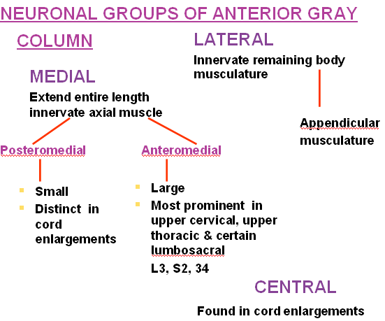
• Four columns of motor neurons can be identified within this lamina;
anterior, ventromedial, ventrolateral, dorsolateral & central each has characteristic dendritic features
Ventral gray columns in lamina IX have somatotopic arrangement:
– Medial areas innervate the axial musculature
– Lateral areas innervate the limbs muscles
PHRENIC NUCLEUS
- The phrenic nucleus is located in the ventromedial area of the ventral horn in C2-C5 segments.
- This nucleus is responsible for the innervation of the diaphragm
SPINAL ACCESSORY NUCLEUS
- The spinal accessory nucleus (cranial nerve XI) is located in the lateral area of vental horn in C1-C5 segments.
- This nucleus is also responsible for the innervation of the trapezius & sternocleidomastoid muscles (ipsilaterally)
Nucleus of Onuf
- Located ventrolaterally in S1-S2 spinal segments
- Supplies muscles of the pelvic floor, including striated muscle sphincters for urinary and fecal continence
Lamina X
- Surrounds the central canal, and includes the ventral gray commissure
- Contains relatively small neurons, radial neuroglia cells & decussating axons
- Some dorsal root afferents terminate here
Histological slides are available here.
 howMed Know Yourself


thanks for sharing the info.that is interesting.
excellent