A large number of normal intestinal flora is greatly influenced by diet. The microorganisms which form part of this normal flora include:
- Coliform bacilli
- Species of Proteus, Pseudomonas, Clostridium, Bacteroides, Enterococcus and Lactobacilli are common
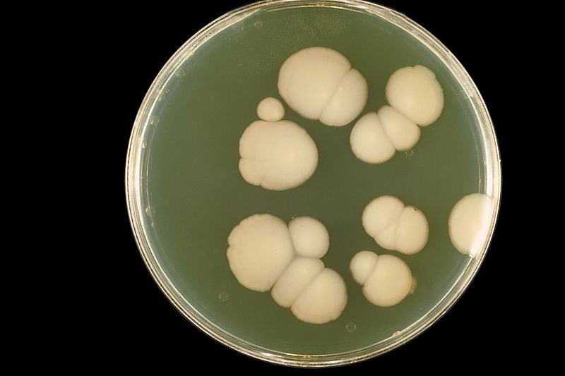
- Mycoplasma, Candida species and a variety of Protozoa may also be present
 Pathogenesis
Gram Positive Bacteria
- Clostridium perfringens type A and C
- Clostridium difficile
- Bacillus Cereus (toxin)
- Staphylococcus aureus (toxin)
Gram Negative Bacteria
- Shigella species
- Salmonella species
- Campylobacter species
- Yersinia enterocolitica
- E. coli
- Vibrio species
- Mycobacterium tuberculosis
Viruses
- Rota virus
- Norwalk virus
- Adenovirus
- Calicivirus
- Corona virus
Parasites
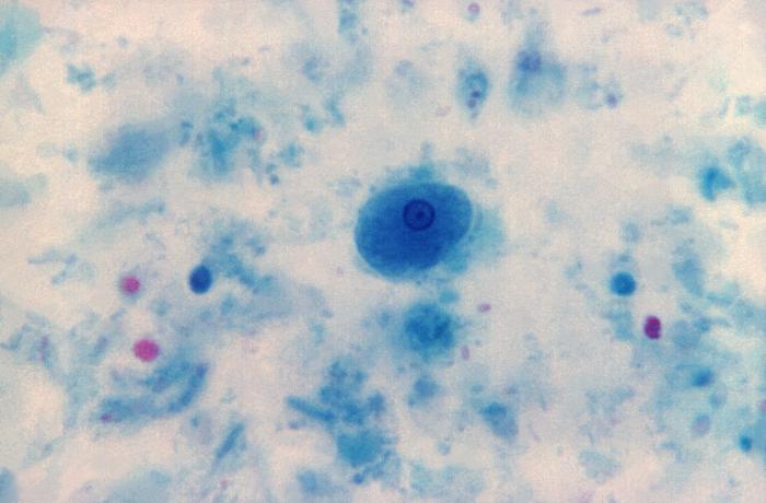
- Entamoeba histolytica
- Giardia lamblia
- Intestinal Coccidia (Isospora, Cryptosporidium, Cyclospora)
- Clostridium perfringens produces type A and C enterotoxins that cause diarrhea whereas Clostridium difficile causes pseudomembranous colitis
- Bacillus Cereus causes diarrhea by preformed toxins in cereals
- Staphylococcus causes diarrhea by preformed toxins in dairy products
- Shigella species cause bacillary dysentery usually in young children
- Salmonella species cause enteric fever and enterocolitis
- The commonest virus that causes diarrhea in young children from 6 months to 3 years of age is Rota virus
- Cryptosporidium is the most common opportunistic parasitic infection causing diarrhea in patients of AIDs.
- Ascaris lumbricoides, Ankylostoma duodenale (hookworm), Giardia and Taenia saginata are common parasites causing diarrhea in humans.
Collection of Sample
Stool sample should be collected in acute phase of diarrhea. A clean, wide neck container should be used. Contamination with urine should be avoided. Label the specimen along with a filled request form and send it to the lab within one hour.
Transport
If delay longer than one hour is anticipated from collection of specimen to submission in lab, the specimen should be taken in Cary Blair medium.
If Salmonella and Shigella are suspected, the specimen should be collected in tetrathionate broth. Similarly, Vibrio cholera should be transported in enrichment medium like alkaline peptone water.
For virus mixed, take 1 ml of sample with 9 ml of phosphate buffer, centrifuge it, and suspended material is sent for examination.
If worm infestation is suspected, the specimen should be transported in saline.
Gross Appearance
Following characteristics are noted:
- Color of specimen
- Whether it is formed, semi-formed or fluid.
- Presence of blood, mucous or pus
- Presence of worms, e.g. Enterobius vermicularis, Ascaris lumbricoides or tape worm segments
- If stool is black in color, there is a chance of upper GI bleeding. Frank blood (fresh) in the stool gives an indication of lower GI bleeding and hemorrhoids.
Microscopic Examination
Examine the specimen on slide. Take 1 drop of specimen and mix it with 1 drop of saline on a slide and emulsify it. Place a cover slip and observe under microscope.
The cysts of Protozoa are better seen when mixed with a drop of eosin. Staining with methylene blue is done for pus cells. if Vibrio cholera is suspected, its motility (shooting star) is observed by dark field microscopy. Shigella is non-motile diarrhea causing pathogen.

I need PDF for stool and urine examination