Chronic granulomatous inflammation is a distinctive pattern of chronic inflammatory reaction in which the predominant cell type is an activated macrophage with modified epitheloid appearance, also called as type IV hypersensitivity reaction or cell mediated immunity.
Mechanism
- Role of T lymphocytes
- Role of macrophages
- Role of giant cells
Granuloma
A granuloma is a focus of chronic inflammation consisting of macrophages transforming to epitheloid cells and surrounded by a collar of mononuclear leukocytes (lymphocytes), and occasional plasma cells. Frequently epitheloid cells join together to form giant cells, either in the periphery or center of granuloma. As it becomes older, there is a rim of enclosing fibroblasts produced along the collar of lymphocytes.
Types of Granuloma
1. Foreign body Granuloma
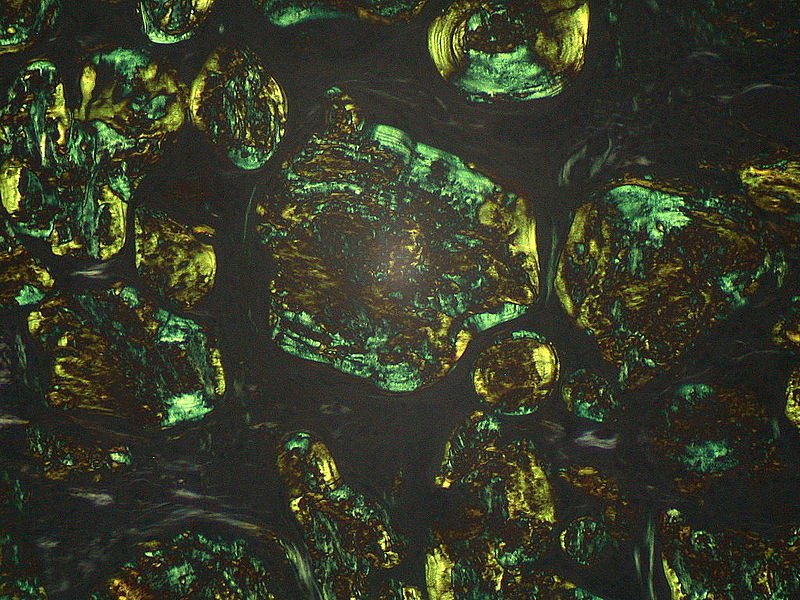
It forms around materials like sutures, talc or other fibers that are large enough to inhibit phagocytosis by a single macrophage, and do not exert any specific inflammatory or immune response. Epitheloid cells and giant cells are opposed to the surface of foreign body, which can be visualized in the center of granuloma, especially with polarized light.
2. Immune Granulomoa
They are caused by variety of agents capable of inducing cell mediated immune response. This type of immune response produces granulomas when inciting agent is poorly degradable. The prototype of immune granuloma is caused by mycobacterium tuberculosis and is referred to as tubercle. It is often characterized by presence of central caseous necrosis, which is rare in other granulomas.
Causes
Infective Causes
Bacteria
- Tuberculosis caused by mycobacterium tuberculosis
- Leprosy by mycobacterium leprae
- Syphilitic granuloma by Treponema pallidum (gumma)
- Cat scratch disease by Bartonella hensalae
Parasitic Causes
- Schiostosmiasis
Fungal Causes
- Histoplasma capsulatum
- Blastomycosis
- Cryptococcus neoformans
- Coccioiodes
Foreign Bodies
Exogenous
- Sutures
- Vascular grafts
Endogenous
- Necrotic bone
- Keratin
- Cholesterol crystals
Malignancy
- Seminoma
- Hodgkin’s lymphoma
Inorganic Metals and Dust
- Silicosis
- Berylliosis
Drugs
- Phenyl butazone
- Sulphonamide
- Allopurinol
Uncommon Causes
- Sarcoidosis
- Crohn’s disease
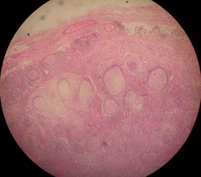
Morphology: Tuberculous Lymph Node
Gross
Any group of lymph nodes can be involved but cervical and axillary are the most common. Mesenteric lymphadenitis occurs in intestinal tuberculosis and enlarged matted lymph nodes with rubbery consistency are common. Calcified lymph nodes are hard and cut section shows greyish white areas with cheesy consistency due to caseous necrosis.

Microscopy
In a typical granuloma there is central caseation surrounded by a zone of epitheloid cells, which have pale, eosinophilic cytoplasm and large vesicular nuclei. There may be scattered giant cells. Nuclei of giant cells are arranged in form of horse shoe and are called Laghan giant cells. In the periphery, there is a zone of mononuclear infiltration composed of lymphocytes and occasional plasma cells and fibroblasts.
Want a clearer concept, also see all images on Chronic Granulomatous Inflammation
 howMed Know Yourself


