►Involuntary muscle
►Innervated by autonomic nerves
►Lack striated pattern (smooth)
Types
►Single unit (visceral smooth muscle)
►Multi unit smooth muscle
Single unit smooth muscle
►Fibers connected by gap junctions
►Function as a single unit (syncytium)
►Stimulated by
►Nervous stimuli
►Non-nervous stimuli (chemicals or physical)
►Present in walls of hollow viscera
►GIT, reproductive & urinary tracts and small blood vessels
Multi unit smooth muscle
►Each fiber functions independently
►Separate nerves to each fibers
►Stimulated by nervous signals only
►Present in
►Ciliary muscle, iris, piloerector muscle
Smooth muscle fiber
►Spindle shaped cell
►Single nucleus
►Smaller than skeletal muscle fiber
►No ‘T’ tubules -caveolae present
►Calmodulin in place of troponin
►Have capability to divide (unlike skeletal muscle)
Contractile unit
►Actin filaments -attached to dense bodies
►Lack troponin
►Myosin filaments
►Single filament within a ‘sarcomere’
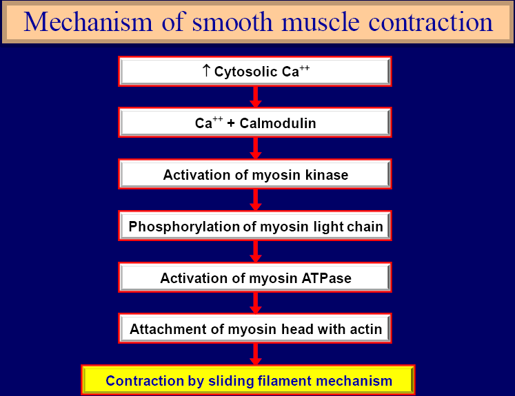
Contraction of smooth muscle
►Ca++performs two functions
►Generation of action potential
►Initiation of contraction
Relaxation of smooth muscle
►Role of myosin phosphatase
►Dephosphorylates myosin head
►Removal of Ca++
►Ca++pump located at fiber membrane
►Dissociation of Ca++-Calmodulin complex
Contraction of smooth muscle
Source of Ca++
►ECF
►Some from sarcoplasmic reticulum (role of caveolae)
Characteristics of smooth muscle contraction
►Less ATPase activity of myosin head
►Slow cycling of myosin cross bridges
►Decreased energy requirement
►Slow onset of contraction & relaxation
►Increased force of muscle contraction
Stress relaxation
►When stretch increases
►Contractile filaments adjust to new position to undo effect of rise in stretch
►Rise in BP, filling of urinary bladder
►Reverse stress relaxation
►When stretch decreases
►Contractile filaments adjust to new position to undo effect of decrease in BP
►Fall in BP
Latch mechanism
►Sustained contraction with little excitatory signal (exactly opposite to skeletal muscle)
►Less use of energy
►Myosin heads remain attached (latched) with actin for much longer time
Electrical activity
►In single unit smooth muscle
►Recording from many muscle cells
►In multi unit smooth muscle fiber
►Action potential can not be recorded from a single cell
Action potential in smooth muscle
Resting membrane potential
►-50 to -60 mv (unstable)
►Depolarization
►Opening of voltage gated Ca++channels
►Repolarization
►Opening of voltage gated K+channels
►Spike potentials
►Action potentials with plateau
►Action potentials on slow waves
Spike potentials
►Typical action potential
►Due to Ca++influx and K+efflux
Action potential with plateau
►Due to Ca++influx and delayed K+efflux
►Leads to sustained contractions
►Occurs in ureters, uterus etc
Slow waves (pacemaker waves)
►Waxing and waning of Na+-K+pump
►At threshold action potential initiates
►Occurs in GIT
Stimuli
►Nervous
►Non-nervous
►Hormones
►Paracrine factors
►NO, prostacyclin, adenosine etc
►Physical
►Stretch
Smooth muscle cell membrane contains receptors
►Receptors either excite or inhibit smooth muscle
Nervous stimuli
►Autonomic nerves
►Make diffuse junctions over sheet of single unit smooth muscles
►Either contract or relax smooth muscle
►Contact junctions with multi unit smooth muscle fibers
Diffuse junctions (with single unit smooth muscle cells)
►No direct contact of autonomic nerves with muscle cells
►Nerves terminate on sheet of cells
►Nerves form varicosities
►Non-nervous stimuli
►Hormones and paracrine factors
►Action through ion channels
►Opening of Na+or Ca++channels (stimulation)
►Opening of K+channels (inhibition)
►Action through 2ndmessenger system
►Stimulation or inhibition of smooth muscle
2ndmessenger system Relaxation of vascular smooth muscle by NO
Non-nervous stimuli
►Physical stretch
►Contraction of smooth muscle
►Bulk laxatives (fiber diet)
 howMed Know Yourself


the presentation iz jst superb, quite easy 2 understand.
student.physical education