Introduction
‘Connective tissue’ is the term traditionally applied to a basic type of tissue of mesodermal origin which provides structural and metabolic support for other tissues and organs throughout the body, also known as “Supporting tissue”.
Functions
• Establishing a structural framework
• Supporting, surrounding and interconnecting tissues
• Transporting fluids and dissolved materials between cells & their blood supply
• Storing energy reserves
• Defending the body from microorganisms
• Protecting delicate organs
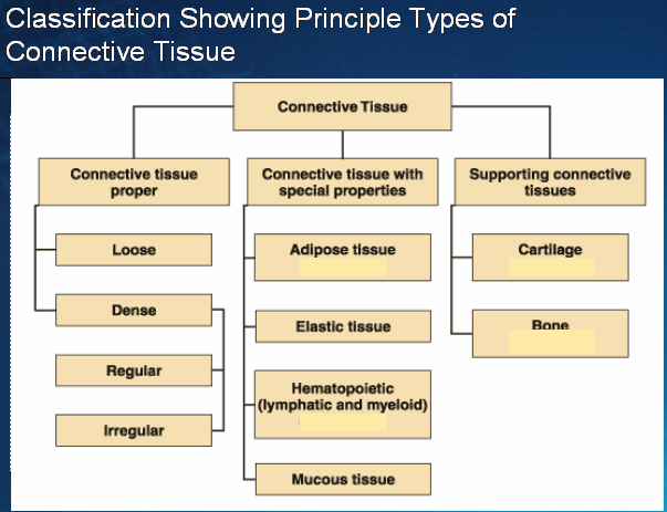
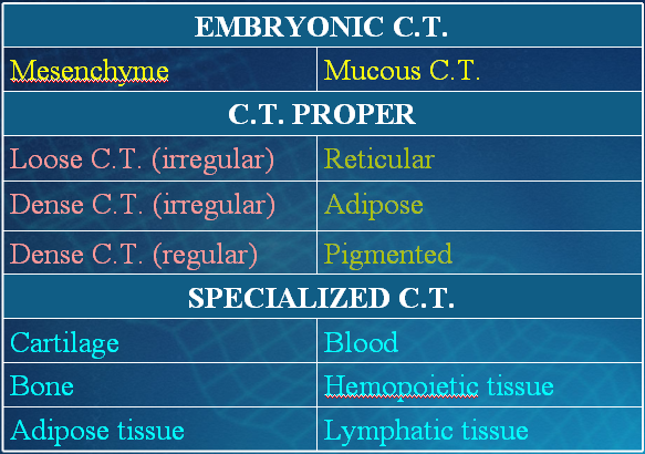
Connecting tissue is broadly classified in to
1) Connective tissue proper
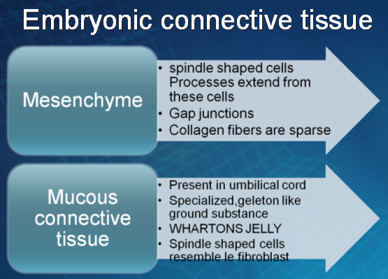
2) Embryonic connective tissue
Composition
- Cells
- Extracellular Matrix
n Fibers
n Ground substance
Cells
v Residents
- Fibroblasts
- Macrophages
- Reticular cells
- Mesenchymal cells
v Visitants
- Mast cells
- Plasma cells
- Leukocytes
- Fat cells
- Melanocytes
Extra-cellular Matrix
• Fibers
-Collagen fibers
-Reticular fibers
-Elastic fibers
Ground Substance
-Proteoglycans
-Glycosaminoglycans
-Glycoproteins
Resident Cells
1. FIBROBLASTS
One of the two most numerous cells of areolar C.T.
Large, flat cells with branching processes and appear fusiform in profile.
Active fibroblasts
Mature/inactive fibroblasts
Most common cells
Synthesize proteins, such as collagen and elastin that forms collagen, reticular, and elastic fibers
Secrete “Ground Substance”
Involved in production of “Growth Factors”
Wound healing
Functions Of Fibroblasts
Fibers producing cells.
Recent research suggests that a single fibroblast is capable of producing all of the extra cellular matrix components
They can change in to myofibroblasts which are rich in actin filaments and help in approximation of wound margins during healing. Myofibroblasts display properties of both fibroblasts & smooth muscle cells
2. Macrophages (Histiocytes)
Almost as numerous as fibroblasts
Most abundant in richly vascularized areas
Fixed/resting macrophages
Free/wandering macrophages
In resting phase
Appear as irregular cells with short and blunt processes but sometimes the processes may be long, slender and branching.
Nucleus is rounder, smaller and more heterochromatic than that of fibroblast.
Cytoplasm stains darkly & may contain few small vacuoles, stain supravitally with neutral red
In active state
• Cell becomes larger with bigger nucleus and prominent nucleolus
• Cytoplasm becomes filled with granules and vacuoles, containing ingested material
• Size 10-30 µm
• Oval or kidney shaped nucleus located eccentrically
• Monocytes-Marophages
• Kupffer cells-liver, microglial cells-CNS, osteoclasts- bone tissue
• Phagocytosis
• Have well-developed Golgi complex, many lysosomes, and a prominent rough endoplasmic reticulum
• ‘Foreignbody giant cell’
Functions Of Macrophages:
Important agent of defense and part of Mononuclear phagocyte system.
These are secretory cells that produces several important substances including enzymes, two proteins of complement system, an important antiviral agent, interferon
They play very important role in immune system and act as antigen presenting cells.
They are capable of motility and when suitably stimulated grouped around a large foreign body to form multinucleated foreign body giant cells
3. ADIPOSE CELLS
The large mesenchymal cells after accumulation of fat droplets in their cytoplasm are called adipocytes
Occur singly or in clumps along small blood vessels
Also called “Fat Cells”
Size 50-150 µm
Cytoplasm is dispalced to periphery by a single large fat droplet
Nucleus is pressed against the cell membrane “Signet ring”
Functions in energy reserves, insulation, protection, and support
Under L/M:
Prior to the storage of fat they are stellate shape cells and difficult to distinguish from fibroblasts but after storage of fat they become rounded.
Consists of peripheral rim of cytoplasm in which oval nucleus compressed against the cell wall is present.
4. Mast cells
Mast means well fed. Their cytoplasm is full of coarse granules so this name is given.
Tend to occur in small groups around blood vessels.
Irregularly oval in outline, have short pseudopodia
Nucleus is small being crowded by large number of prominent granules
Oval/round, Size 10–13 µm
• Secretory granules (0.3-2 µm)
• Histamine & Heparin
• Metachromatic granules
• Two populations of mast cells in CT
• ‘Connective tissue mast cells’ (skin, peritoneal cavities)-heparin
• ‘Mucosal mast cells’ (intestinal mucosa, lungs)-chondroitin sulfate
Functions Of Mast Cells
As these granules contain heparin (a powerful anticoagulant) and histamine (a potent vasodilator) so they play very vital role in homeostasis.
• Mast cells are especially numerous in C.T. of skin & mucous membranes, but are not present in brain & spinal cord. These are numerous in thymus, & to a lesser degree, in other lymphatic organs, but they are not present in spleen.
The release of chemical mediators (such as ECF-A), Leukotriene C, PAF by the mast cells against antigens promotes the allergic reaction known as immediate hypersensitivity reaction.
5. PERICYTES
• These are cells which are present external to endothelium of blood capillaries and small venule.
• Cytoplasm is pale staining and possesses long processes.
• They can differentiate into fibroblasts, smooth muscle cells and myofibroblasts during wound healing.
Wandering Cells
1. LYMPHOCYTES
Smallest of the free cells of C.T.
Majority being 7-8µm in diameter
Spherical darkly staining nucleus
Thin rim of homogenous basophilic cytoplasm
Numerous in C.T. that supports epith lining of respiratory and elementary tracts
2. PLASMA CELLS
Resemble to lymphocytes
More cytoplasm which is basophilic
Eccentric nucleus
Cartwheel appearance
Cytoplasm contains a clear, rounded area (site of centrosphere and golgi apparatus
Extensive RER
Frequently found in serous membranes and lymphoid tissue and plentiful in sites of chronic inflammation
Represent a special differentiation of lymphocyte
Principle function is the production of antibodies which are synthesized in RER
Acidophilic inclusions called Russel bodies are present in cytoplasm
Large, ovoid cells
Nucleus is spherical and eccentrically placed
‘cart-wheel appearance’
Basophilic cytoplasm due to abundant RER
Rare in most connective tissues
Produce antibodies
Average life is short, 10–20 days
3. EOSINOPHILS
Not numerous in human C.T.
Plentiful in C.T. of lactating breast and of respiratory and alimentary tracts
Marked feature of loose C.T. of rat, mouse and guinae pig
Nucleus is usually reniform or bilobed
Cytoplasm contains spherical granules that are highly refractile and stain with acid dyes
Accumulate in blood and tissues in certain allergic and subacute inflammatory conditions
Wandering cells
leukocytes that migrate from the blood vessels by diapedesis
This process increases greatly during inflammation
Fibers
• Formed by proteins which polymerize into elongated structures
• Three types
• Collagen Fibers
• Reticular Fibers
• Elastic Fibers
• Collagen and reticular fibers are formed by the protein collagen, and elastic fibers are composed mainly of the protein elastin
Collagen
- Family of closely related proteins
- Composed of long molecules (tropocollagen)
- Each molecule is 280 nm in length & 1.5 nm in width and consists of three polypeptide chains in the form of right handed triple helix
- 14 varieties of collagen, type I – type XIV collagen
Collagen Fibers
• Most abundant protein in human body
• 30% of body weight
• Made up of collagen type-1
• Diameter is variable(2-10 µm) and have great indefinite lengths
• Tough and flexible but in-elastic (non-extensible)
• EM shows that each collagen fiber is composed of parallel fibrils (50-90 nm in diameter) which appear to be cross striated
Reticular Fibers
• Mainly consist of type III collagen
• Extremely thin fibers (diameter 0.5-2 µm)
• Form extensive network
• Not visible by H&E stain
• stained by impregnation with silver salts (argyrophilic)
• PAS positive
• Abundant in smooth muscle, endoneurium, hemopoetic organs (spleen, lymph nodes, bone marrow)
Elastic Fiber System
• Three types of fibers—oxytalan, elaunin, and elastic
• Oxytalanic fibers (zonule fibers of eye) contain
• ‘fibromodulin I and II’ and ‘fibrillin’
• Elaunin fibers (sweat glands, dermis)
• Elastic fibers (lungs, large arteries, ligamentum flavum)
Ground Substance
- Fine granular material
- Fills the space between cells & fibers of CT
- Highly hydrated, colorless & transparent
- Formed by three components
- Proteoglycans
- Glycosaminoglycans
- Multiadhesive Glycoproteins
- a. Fibrnectin
- b. Laminin

-
Need to clear your concept? Also see
-
Types of Connective Tissues
-
Histological slides of loose connective tissue
-
Histological slides of adipose tissue
-
Histological slides of dense connective tissue
 howMed Know Yourself


