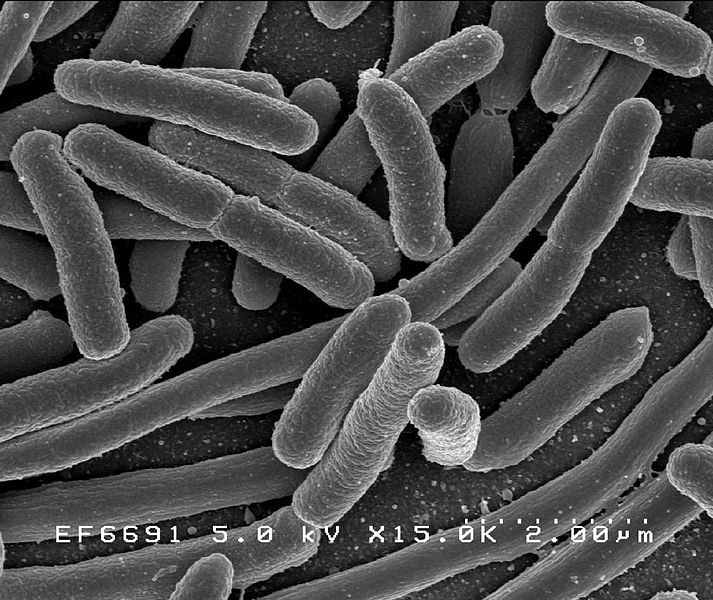
Escherichia coli is a gram negative bacteria, rod shaped and facultative anaerobic.
Route of entry
• Most abundant facultative anaerobe in the colon and feces
• Food or water contaminated with human feces
• Undercooked meat
Pathogenesis
E.coli has 3 antigens:
a. O cell wall antigens
b. H antigens
c. K antigens
Enterotoxigenic Escherichia coli (ETEC)
Causes watery diarrhea to production of plasmid meditated toxins, ST and LT
Enteropathogenic Escherichia coli (EPEC)
Causes vomiting, fever and prolonged diarrhea in infants due to bacteria adhering to epithelial cells, multiplying and causing lesions
Enteroinvasive Escherichia coli (EIEC)
Causes dysentery, fever and colitis with blood mucus and many pus cells in the fecal specimens due to bacterial invading and multiplying in epithelial cells.
Enterohemorrhagic Escherichia coli (EHEC)
Causes life threatening hemorrhagic diarrhea in all ages, without pus cells and often without fever. Due to cytotoxins damaging vascular epithelial cells, mainly toxin can cause hemolytic uremic syndrome consisting of anemia, thrombocytopenia and acute renal failure.
Enteroaggregative E. coli (EAEC)
Causes chronic watery diarrhea and vomiting mainly in children due to bacteria adhering to tissue cells. The capsule and the endotoxins play an important role in systemic infections. Urinary tract infections are caused by uropathic strains, characterized by pilli.
Virulence factors
a. Pili, protrude from the bacterial surface and aid attachment on host cell membrane
b. Capsule, anti-phagocytic properties
c. LT, catalizes the addition of adenosine diphosphate ribose to G-Protein
d. ST, stimulates guanylate cyclase
e. SLT, removes an adenine from the large ribosomal RNA, therefore stopping protein synthesis
f. Endotoxins, causing fever and shocks
Clinical symptoms
• Community acquired UTI mainly in women
• Nosocomial UTI
• Cystitis
• Pyelonephritis
• Meningitis
• Peritonitis
• Diarrhea
• Travelers’ diarrhea
• Bloody diarrhea
Lab Diagnosis
Specimen
Specimen depends on infected site:
• urine
• pus
• feces
• CSF
• blood
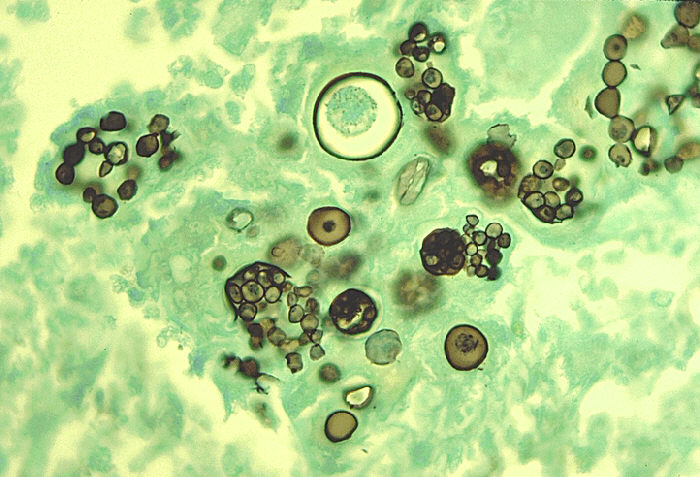
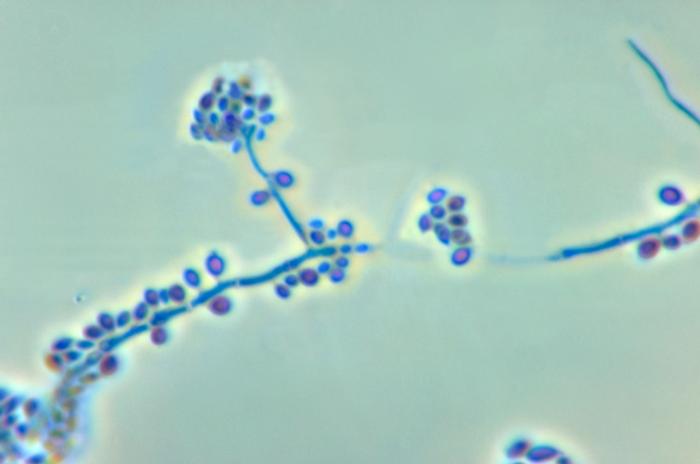
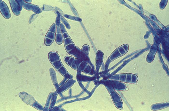
Microscopy
• Straight gram negative rods
• Motile
• Facultative anaerobes
• Enapsulated
• Pili are present
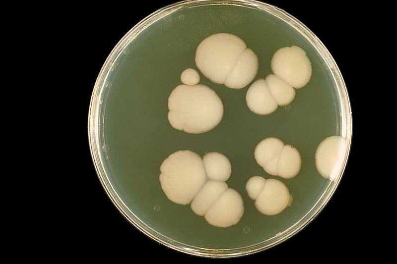
Culture
Blood agar: mucoid colonies. Some strains are hemolytic
MacConkey agar: smooth yellow pink colonies
CLED: yellow colonies
XLD agar: yellow colonies
DCA agar: growth is inhibited
Biochemical Tests
• catalase positive
• oxidase negative
• indole positive
• H2S negative
• citrate negative
• B-Glucuronidase positive
Serological Test
PCR
Treatment
Treatment depends on site of disease and resistance pattern of organism.
• ORS
• Penicillin