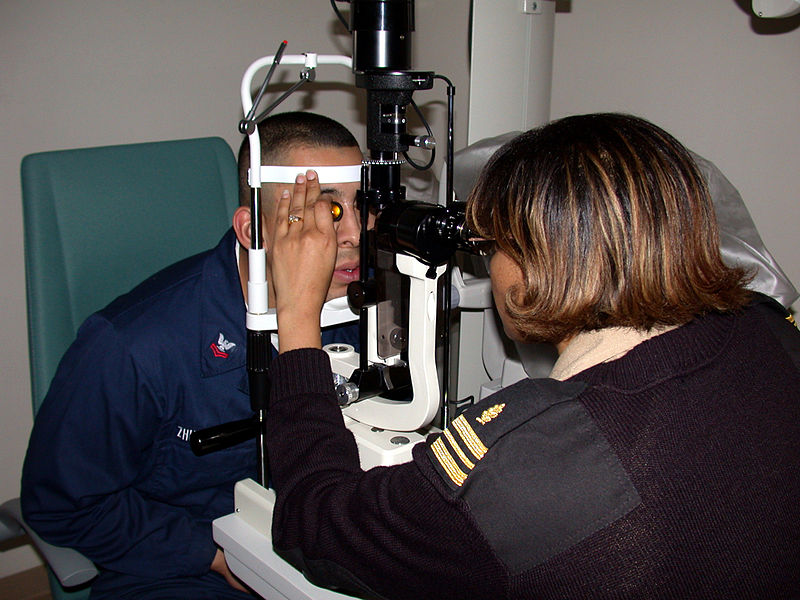
The steps involved in examination of posterior segment of eye may be summarized as:
- Introduction
- Visual acuity checked grossly
- Check settings:
- High illumination
- High magnification
- Slit beam
- Check patients stool height place patient face and adjust lateral canthus.
- Examine anterior vitreous face setting for counting cells
- Check setting (90D)
- Medium illumination
- Low magnification
- Slit beam
- Examine:
- Posterior hyaloids face of vitreous
- Disc, Macula, Vessels.
- For periphery ask patient to look in all direction of gazes
- Check Setting:
- Red free filter
- Examine:
- Posterior hyaloids face
- Nerve fiber layer
- Vascular pathology
- I would like to examine with triple mirror and indirect 0phthalmoscope (if pathology or absolutely normal)
- Thank the patient and make him sit back and turn off the slit lamp.

 howMed Know Yourself


