Coagulative necrosis is the most common pattern of necrosis characterized by denaturation of cytoplasmic proteins, cellular swelling and breakdown of cellular organelles. The most common organs involved are heart, kidney and spleen.

Morphology
Gross
In heart, in case of coagulative necrosis, the tissue appears hard, dry, firm and white. The cellular outline and tissue architecture is well preserved. Up till half an hour, the injury is reversible and there is no visible changes in myocardium up to two to three hours. We can highlight the area of necrosis by putting a dye i.e. triphenyl tetrazolium chloride. This dye imparts brick red color to non-infarcted area. The area of coagulative necrosis becomes prominent as pale zone.
Microscopic
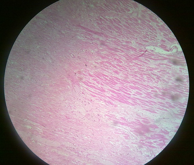
Typical microscopic changes in coagulative necrosis become prominent in 4-12 hours, there is preservation of basic outline of injured cells along with denaturation of cellular proteins. There is increased eosinophilia due to denaturation of proteins, loss of normal basophilia imparted by RNA degeneration. Secondly, eosin stain has more affinity for denatured proteins. Some nuclear changes take place in myocardial fibers. Normal striations are lost due to necrotic changes, but in some areas myocardial fibers may show striations, which is due to reperfusion of that area. Interstitial neutrophilic infiltrate is seen in between the dead muscle fibers. These neutrophils come for phagocytic action to remove the myocardial cells which have become necrotic.
 howMed Know Yourself


