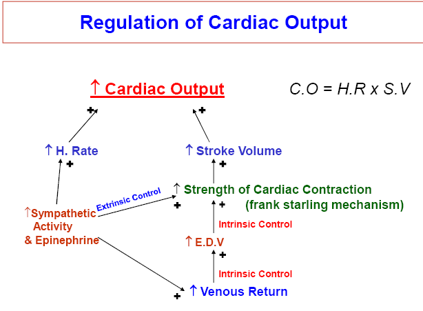
Quantity of blood pumped into aorta each minute by the heart.
C.O approx.= Venous Return.
C.O = H. Rate x Stroke Vol.
= (70/min) x (80 ml/beat)
Cardiac output is also called ‘minute volume’.
Normal range of C.O = 5-7 L/min
Stroke vol: (S.V)
Amount of blood pumped out of each ventricle per
beat; also called ‘Systolic discharge’.
S.V = E.D.V – E.S.V
= 140 ml – 60 ml
S.V = 80 ml/beat in resting man.
Cardiac Index:
C.O per minute per square meter body surface area.
C.I = C.O/S.A = 5.0/1.7
C.I = 3.2 L/min/m2
Stroke Vol Index:
S.V per square meter body surface area.
S.V.I = 47 ml/m2
Preload and Afterload
Preload:
Vol of blood in the ventricle at the end of diastole (E.D.V)
Degree of tension on the muscle when it begins to contract is called
preload.
Preload is also the degree of filling of ventricle and depends on the
venous return.
Factors affecting preload:
Posture
Bl.vol
MSFP
Atrial contraction
Muscular activity
Intrapleural pr.
Intrapericardial pr.
Frank Starling Law:
Force of contraction is proportional to the initial length of
cardiac muscle fiber (i-e preload or E.D.V).
Increased Preload or E.D.V leads to
1)Increased Force of contraction by Frank Starling Law
2)Increased Heart Rate (Approx. 75%) by
a)direct stretch on S.A Node (10-15%)
b) by Bainbridge Reflex or Cardioaccelerator Reflex (40-60%)
After load:
Arterial pressure against which ventricle contracts
i.e.
Systolic pressure, Peripheral resistance or Impedance or resistance.
Thus; increased Afterload = decreased Cardiac output
I. Increased:
Eating (30%)
Anxiety/Excitement (50-100%)
Exercise (up to 700% i-e 5-7 fold)
High environmental temp.
Pregnancy
Epinephrine, Histamin
Anemia
Metabolic Alkalosis, Low PO2 or High PCO2.
Age up to puberty
II. Decreased:
Sitting or standing from supine position (20-30%)
Rapid arrhythmias
Heart diseases (Hypo effective Heart)
III. No Change:
During sleep
Moderate change in environmental temp.
Metabolic acidosis
Menstruation
However, C.O is regulated throughout life directly in
proportion to the metabolic activity.
High Cardiac Output:
Always caused by decreased Total peripheral resistance.
1. Beri Beri
2. A-V fistula/shunt
3. Hyperthyroidism (increased Metabolism, increased Vasodilator subs= decreased T.P.R)
4. Anemia. (decreased Bood Viscosity, Hypoxia leads to decreased T.P.R)
Low Cardiac Output:
1. Decreased pumping effectiveness of heart:
Severe myocardial infarction
Severe valvular heart disease
Myocarditis
Cardiac temponade.
2. Decreased Venous Return (decreased EDV):
Blood Volume
Acute venous dilatation (Sudden inactivity of
sympathetic system).
Obstruction to large veins (Rare)
Measurement of Cardiac Output:
1) Direct Method:
In animals
By direct cannulation of Aorta or Pul. Artery
and by flowmeters.
2) Indirect Method:
In Human beings, No surgery required.
i. Fick ‘s Method O2 Fick Method
ii. Dye Dilution Method
iii. Flowmeters (Ultrasonic or Electromagnetic)
iv. Doppler Method.
O2 Fick Method:
Fick Principle:
Amount of substance taken up by an organ (or by whole
body) per unit time is equal to the arterial level of substance
minus the venous level (AV difference) times the blood flow.
In O2 Fick method; C.O is measured by determining the
amount of O2 consumed by the body in a period of time and
dividing this value by A-V difference across lungs.
 howMed Know Yourself


Medicine in general is quite interesting. It explores and explains reasons of one being human.