Heart is innervated by
both divisions of autonomic nervous system (ANS)
i.e. sympathetic &
parasympathetic.
• Nervous stimulation is not required to initiate cardiac impulse.
• ANS Can modify rate, rhythm & force of contraction.
ANS Can modify rate, rhythm & force of contraction.
Parasympathetic nerves to heart:
– Through Rt Vagus (to SA node) & Lt Vagus (to AV node) Nerves
–Main supply to SA & AV nodes but less to Atria & very little to ventricles
– Vagal fibers (Cholinergic) are endocardial
Sympathetic nerves to heart:
– Through Rt & Lt Stellate ganglions
– SA node, AV node & Ventricular muscles
– Noradrenergic fibers are epicardial
Reciprocal Inhibition by Sympathetic & Parasympathetic
innervations to heart:
– Ach acts presynaptically to inhibit NE release from sympatheic nerves
– Neuropeptide-Y from noradrenergic endings inhibits Ach release
Role of Autonomic Nerves
• Parasympathetic:
– Controls heart action in quiet, relaxed situations;
when body is not demanding enhanced CO.
– Rt Vagus —– Slows heart ( ↓ Heart Rate)
– Lt Vagus —– Slows AV conduction (↑ AV nodal delay)
• Weak to moderate stimulation: ↓ heart pumping by 50%
• Strong stimulation abolish spontaneous discharge; ventricular escape
Mechanism of Action of Ach:
• Mediated by M2 muscarinic receptors via βγ subunit of G protein &
opens special K+ channels i.e. ↑ Pk+ (↑K+ conductance)
(Hyper polarization)
• M2 receptors decrease cAMP in cells, slows opening of Ca++ channels,
decreases firing rate (↓Heart Rate)
Sympathetic:
– Controls heart in emergency or exercise
– Rt stellate ganglion → SA node → Accelerates heart through pacemaker
– Lt stellate ganglion → ↓AV nodal conduction time& refractoriness → →
→ ↓ AV nodal delay → → Accelerates conduction
• Strong stimulation → ↑ overall activity of heart
• H.Rate 3 x times
• Strength 2 x times
Mechanism: NE binds β1 receptors → ↑ intracellular cAMP → →
→ opens L-type Ca++ channels → membrane potential
fall rapidly (depolarization)
Effects: ↑ Rate of Discharge (Chronotropic)
↑ Rate of conduction (Dromotropic)
↑ Level of excitability (Bathmotropic)
↑ Force of contraction (Inotropic)
Regulation of Heart Pumping
‘Regulation of heart rate and contractility’
• At Rest heart pumps 4 – 6 L of blood per min
• During strenuous exercise : 4 -7 folds increase.
Regulating Mechanisms:
1. Intrinsic cardiac regulation;
“Frank- Starling’s Mechanism”
2. Role of ANS;
• Sympathetic
• Parasympathetic (Vagus)
3. Other mechanisms;
• Hormones; Catecholamines; Epi & NE
Thyroxin
• Ions; K+
Ca++
• Temperature; Fever (10 beats/min/1◦F rise)
Hypothermia (60-70 ◦F)
• Age
• Gender
• Physical Fitness “Athletes Heart”
Frank – Starling’s Mechanism
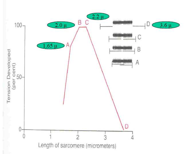
• Intrinsic ability of heart to adapt to the changing volumes EDV
• i.e. greater the heart muscle is stretched during filling (EDV), the
greater will be the force of contraction and greater the amount of
blood pumped out.
• Definition:
“Within physiological limits, the heart pumps all the blood that
comes to it without allowing excessive damming of blood in veins”
• Mechanism:
↑ EDV cardiac muscle stretched so ↑force of contraction
Optimal degree of interdigitation of Actin & Myosin filaments
↑Venous return leads to stretching of Rt atrium ↑H.Rate by 10-20%
Activation of SA node (Bain- Bridge Reflex)
 howMed Know Yourself

