Origin & Propagation Of Cardiac Impulse
Carried out by:
• Specialized Excitatory & Conducting system
• Makes 1% of cardiac cells
Auto rhythmic cells & Conducting cells:
1. S-A Node
2. Interatrial & Internodal pathways
3. A-V Node
4. Bundle of His } Purkinje cells
5. Right & Left bundle branches } Purkinje cells
Sino- Atrial Node
Small, flattened, ellipsoid strip of specialized cardiac muscle
• 3 mm wide, 15 mm long & 1 mm thick
• Located in superior lateral wall of Rt Atrium below & lateral to the opening of superior vena cava
• Smaller (3-5μ) in diameter than atrial muscles (10-15μ)
• Cells are connected through gap junctions
• Called ‘P’ cells i.e. pacemaker of the heart
Interatrial Pathways
• Extend from S.A node within Rt atrium to Lt atrium
• Comprised of small bundles of atrial fibers
• Largest is ‘Anterior Interatrial band’
(from Ant. wall of Rt atrium to left atrium).
• Velocity of conduction 1 m/sec
Inter nodal Pathways
• Extend from SA node to AV node
• Contain purkinje type of fibers
• Connected to A.V node through very small (Transitional) fibers
• Three bundles of atrial fibers:-
1. Anterior internodal tract of Bachman
2. Middle internodal tract of Wenchebach
3. Posterior internodal tract of Thorel
AV Node
Located in posterior septal wall of Rt atrium beneath endocardium (behind the tricuspid valve, adjacent to the opening of coronary sinus)
• The only point of electrical contact between atria and ventricles
• Slow conduction velocity
• Connected to Bundle of HIS through penetrating portion of AV bundle
Bundle of HIS
• Comprised of purkinje fibers (even larger than ventricular fibers)
• Connect penetrating portion of AV bundle to ventricles.
• Connected through gap junctions
Rt & Lt Bundle Branches
• Comprised of Purkinje fibers
• Distal portion of AV bundle divides (5-15 mm towards apex) into Rt & Lt branches
• Located beneath endocardium, progressively divides into smaller branches
Generation of Cardiac Impulse
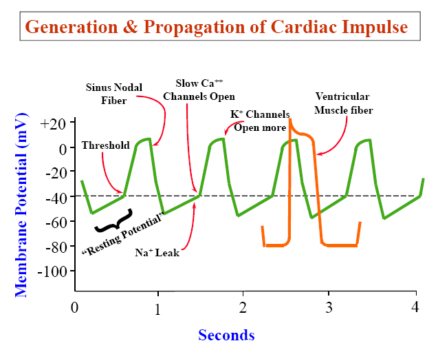
Self excitation/Pace maker potential
• Cardiac cells capable of auto rhythmicity or self excitation are:-
1. SA node 70-80/min
2. AV Node 40-60/min
3. Bundle of HIS 15-40/min
• SA node being the fastest in generation of AP, therefore acts as ‘pace maker’ of the heart.
• Cardiac impulse: is the ‘action potential’ generated in the ‘cardiac pace maker’ and transmitted to the rest of the ‘heart’.
Pace Maker Potential
Mechanism of Self Excitation:
• Cardiac auto rhythmic cells don’t have resting potential.
• Membrane potential slowly depolarizes to threshold and AP is initiated.
• Cycles of ‘drift and fire’ cyclically initiate AP to trigger rhythmic beating of the heart.
Phases:
– Initial phase of ‘slow depolarization’
– ‘Threshold potential’
– ‘Rising phase’ of Action Potential
– ‘Falling phase’ of Action
Prepotential
• Prepotentials are normally present in SA & AV nodes
• ‘Latent Pacemakers’ exist in other portions of conducting system (can take over when SA / AV nodes are blocked)
• Atrial & Ventricular muscles don’t have prepotentials
• Atrial & ventricular muscles discharge spontaneously when injured or abnormal
• Membrane potential slowly depolarizes to threshold and AP is initiated.
• Cycles of ‘drift and fire’ cyclically initiate AP to trigger rhythmic beating of the heart.
Phases:
– Initial phase of ‘slowdepolarization’
– ‘Threshold potential’
– ‘Rising phase’ of Action
Potential
– ‘Falling phase’ of Action Potential
Transmission of Cardiac Impulse in Conducting System
1. Transmission through Atria & Internodal Pathways:
SA node to AV node (Initial Delay) = 0.03Sec
2. Transmission Through AV node (AV nodal Delay)
In AV node itself = 0.09 sec
In penetrating portion of AV bundle = 0.04 sec
Total = 0.13 sec
Total delay from SA node to ventricle
muscles = 0.16 sec
i.e. Initial delay & A-V nodal Delay
(0.03 + 0.13 = 0.16 sec)
Reasons of A-V Nodal Delay:
– Small diameter of AV nodal fibers
– Few gap-junctions between muscle cells & conducting pathways
– Slow speed of conduction in Junctional fibers @ 0.3 m/sec
3. Transmission in Ventricular Purkinje system:
Fast speed of conduction @ 1.5 – 4 m/sec
• Very large size fibers (even larger than ventricular muscles)
• Very high level of permeability of Gap-junctions
4. Transmission in Ventricular Muscle:
• Velocity = 0.3 – 0.5 m/sec (1/6th of velocity in Purkinje fibers)
• From endocardial to epicardial surface of ventricles = 0.03 sec
• Total time for transmission of cardiac impulse from initial bundle
branch to the last ventricular muscle fiber = 0.06 sec
Sequence of Depolarization
• Begins at left side of interventricular septum, moves first to Rt across Mid septum
• Apex of heart
• Side wall of both ventricles
(Endocardial to Epicardial surface)
• Posterobasal part of both ventricles, pulmonary conus & upper most portion of septum
• Transmission of cardiac impulse through heart = 0.22 sec
Transmission of Cardiac Impulse through Heart (Summary)
Transmission of cardiac impulse through heart = 0.22 sec
• From SA node (@0.05m/sec) to AV node = 0.03 sec
• Within AV node (@0.05 m/sec) = 0.09 sec
• Penetrating portion of AV Bundle (@ 0.05m/sec) = 0.04 sec
• Bundle branches & Purkinje fibers (@ 4m/sec) = 0.03 sec
• In Ventricular fibers (@ 0.5m/sec) = 0.03 sec
Special Features of Conductive System
1. Self Excitatory; excitation ‘not dependent’ on external stimulus.
2. Different rate of discharge of different auto rhythmic cells.
3. Auto rhythmic cells interconnected through gap Junction
4. S A node is the fastest in impulse generation thus pacemaker of heart.
5. AV nodal delay:
• Makes atria to contract earlier than ventricles
• Makes atria to act as primer pump
6. Makes all portions of ventricles to contract simultaneously for effective pressure generation.
7. Action potentials in conductive system cannot travel backward ———–‘prevents reentry’.
Cardiac Rhythmicity – Applied Physiology
• Autorhythmicity: (Intrinsic Rhythmicity)
• Real pacemaker: SA node : 70-80/Min
• Potential or latent pacemakers: AV node 40-60/min
Purkinje 15-40/min
• SA node overdrives potential pacemakers to keep them suppressed.
Reasons:
a. Rapidly changing membrane potential of SA node
b. Prolong refractory period of latent pacemakers
• Ectopic Pace maker: Any pacemaker other than SA node
1. Irritable focus e.g.: Ischemia, Atheroma, Plaque
2. Sinus block
3. AV Block; Ventricular Escape
Stoke’s Adam syndrome
Cardiac Rhythmicity – Applied Physiology
Stoke’s Adam syndrome:
• Blood supply to AV node is not sufficient
• AV node will not function i.e. no conduction from SA node
• Heart stops (Overdrive suppression) for 5-15 sec or ≥ 1 min
• Fainting / dizziness due to (Ischemia) ↓ blood flow to brain
• Purkinje fibers take 4-5 sec or more to pick up heart beat.
Artificial pacemaker: Implanted device Temporary or Permanent
 howMed Know Yourself


excellent points and the details are more specific than elsewhere, thanks.
– Murk
superbbb
i like it because you made it brief and satisfactory intellectually.keep it up fellows