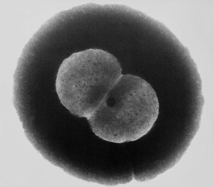
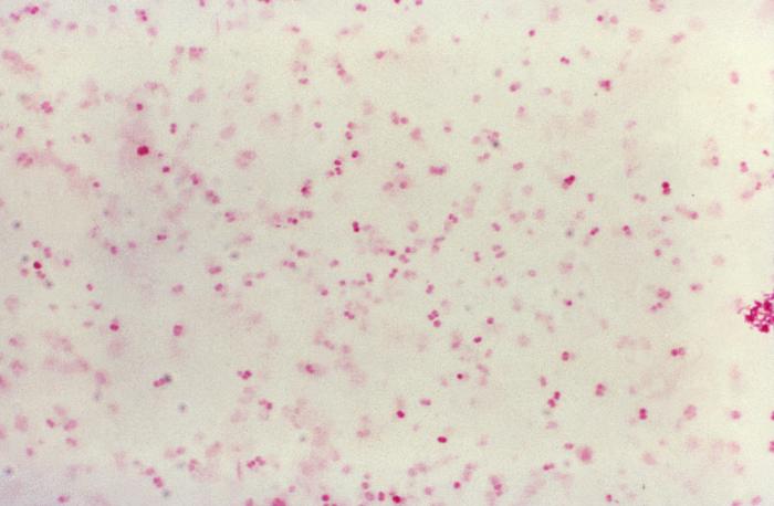
Neisseria gonorrhoeae is a species of gram negative diplococci, which are non encapsulated, non motile and kidney shaped bacteria.
Pathogenesis
Route of entry
Neisseria gonorrhoeae is transmitted sexually. The newborns are infected at birth.
Neisseria gonorrhoeae infects primarily the mucosal membranes. The dissemination occurs through blood stream. Serum resistant bacteria are not killed by anti bodies and complement.
Virulence factors
• Lipooligosacchrides
• Outer membrane proteins porin A
• IgA protease
• Pilli
Lipooligosacchrides
Endotoxin, weaker than lipopolysaccharide, but still causes fever and shock, but to a lesser extent.
Porin A
Inactivates the C3b component of the complement and plays an important role in serum resistance.
IgA protease
Helps bacteria to attach to the membranes by cleaving IgA
Pilli
Mediate attachment to mucosal cell surfaces and are anti phagocytes.
Predisposing Factors
• decrease in secretory antibodies
• deficiency of the late acting complement component
• decrease in neutrophils
• sexually active adults
• during birth, if mother is infected
Symptoms
It causes local and disseminated infections. Gonorrhoea in membranes is characterized by:
• Urethritis
• Dysuria
• Purulent discharge
• Epididymitis
In women cause:
• Purulent discharge
• Cervicitis
• Salpingitis
• PID
• Sterility
Disseminated Gonococcal Infections:
• Arthritis
• Tenosynovitis
• Pustules in the skin
• Anorectal infections mainly in women and homosexual men
• In throat, pharyngitis
• In newborns, gonococcal arthritis
• Vulvovsginitis in pre-pubertal girls
Lab Diagnosis
Specimen
1. Urethral and cervical exudates
2. Urine — male patients
3. Rectal swab when indicated
4. Eye swab
Microscopy
• Gram negative diplococci
• Non encapsulate
• Non motile
• Kidney shaped
Culture
On Thayer Martin medium, small raised grey or translucent colonies are formed in 3-10% CO2.
Biochemical Tests
• Catalase positive
• Oxidase positive
• Ferment glucose only
• DNAase negative
• B-galactosidase negative
• Glutamyl-aminopeptidase negative
Serological tests
- Monoclonal antibody based slide agglutination tests
- PCR
Treatment
Treatment involves ceftriaxone 250mg I/M single dose.