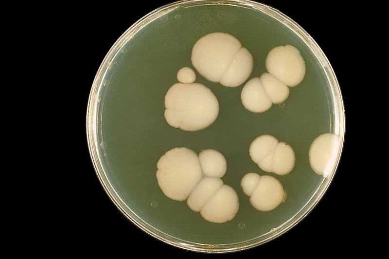
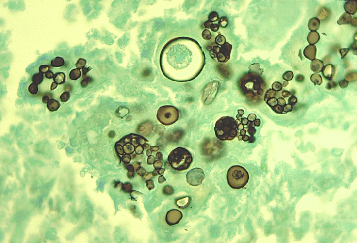
To examine the ovas, cysts or eggs of parasites, take a drop of stool. Mix it with normal saline on a slide and cover the slide with a cover slip. Examine first under low power (10x) and then under high power (40x). Eosin stain can be used as cysts are better seen against pink background.
Protozoa
Entamoeba Histolytica
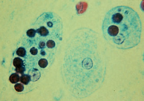
Trophozoite Form
Round to oval in shape, composed of cytoplasm and endoplasm. It contains vacuoles, ingested RBCs and a single nucleus. It is the causative agent of amoebic dysentery and amoebic liver abscess and cause flask shaped ulcers in small intestine.
Cyst Form
Cyst is colorless but refractile. It contains four nuclei and mature chromidial bars. If we put a drop of Lugol’s iodine, it stains nuclei with brown color but chromidial bars remain unstained.
Giardia Lamblia
Its unencysted (flagellated) form is pear shaped with two nuclei, two axostyles and eight flagella in four pairs. It is transmitted by feco-oral route and cause giardiasis.
Cyst Form
It is oval in shape and possess 2-4 nuclei with small fibrils.
Nematodes
Ascaris Lumbricoides
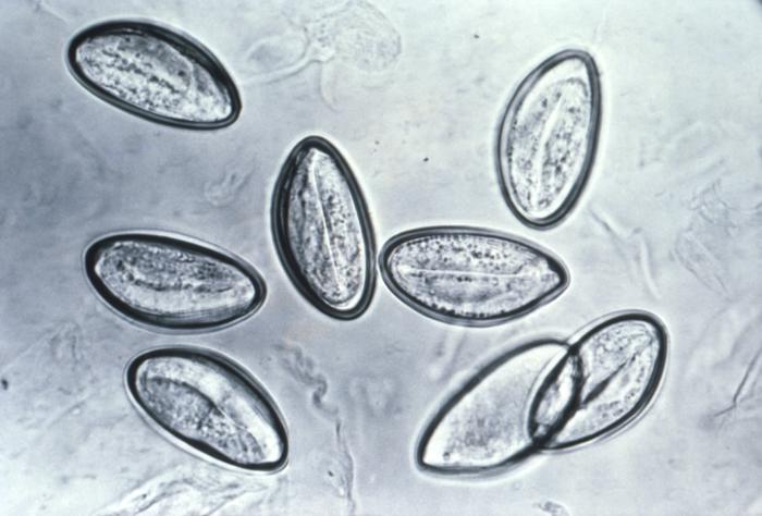
Eggs are round and oval in shape, brownish in color, egg shell is colorless, outside the egg shell is an albuminous coat which is thrown into corrugations.
Ancyclostoma Duodenale
Oval and colorless eggs with thin outer shell, lined by very fine vitelline layer. There is a clear space between egg shell and segmented ovum. In the center, it contains segmented ovum with 2-8 cells.
Trichuris Trichura
The eggs measure about 50 µm in length and 25 µm in breadth. They are brown colored, barrel shaped with a plug at each pole and contains fertilized ovum in the center.
Enterobius Vermicularis
They have colorless eggs, asymmetrical in shape and one side is slightly flattened. Eggs are deposited on perianal skin and not found in stools.
Cestodes
Hymenolepis Nana
Eggs are composed of hexacanth embryo with embryophore and an outer covering. There is a space between outer covering and embryophore which contains polar filaments, 8-10 on each pole of embryo.
Taenia Species
Egg is composed of hexacanth embryo with embryophore.