Amyloidosis is a medical condition resulting from aggregation of extracellularly deposited abnormal proteins called amyloid fibrils that cause damage to organs and tissues. These fibrils are insoluble, linear, rigid and measures approximately 7.5 to 10µm in width.
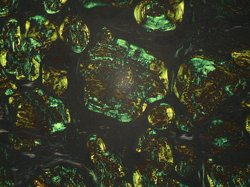
Hematoxylin and eosin stains hyaline acellular eosinophilic material. Congo red stains red in light & green birefingence in polarized light.
Electron microscopy shows regular fibrillar structure. X-ray diffraction shows beta pleated sheet structure.
In 1854 Rudolph Virchow named it amyloid based on color after staining these proteins with iodine and sulfuric acid, amyloid meaning cellulose or starch.
Classificaton of Amyloid
23 different human subtypes have been named based on A for amyloid, followed by the precursor protein e.g AL, AH. Amyloid may be localized or systemic:
Localized
e.g. in larynx, lung or bladder
Systemic Amyloidosis
Systemic amyloidosis may be:
a. Multiple myeloma associated
b. Reactive (secondary amyloidosis)
c. AA amyloid
• Rheumatoid arthritis
• Ischemic bowel disease
• Osteomylitis
• Hodgkins disease & renal cell carcinoma
• Hereditary amyloidosis
Mechanism of Formation
Amyloid fibrils arise from misfolded proteins ranging from alpha helix to beta pleated sheets.
• Proteins are deposited extracellularly
• Proteins aggregate and form fibrils called amyloid fibrils.
• Misfolded proteins may result from point mutations.
• They are deposited as localized vs systemic
– localized; close to cells producing it.
– Systemic; distant sites from these cells producing these abnormal proteins.

Photo courtesy of Ed Uthman, MD under Creative Commons Attribution-Share Alike 2.0 Generic license.
Further Clinical Manifestations
CNS:
– Neuropathy both autonomic and peripheral,
– Dementia.
– Corneal deposits.
Cardiac:
– Cardiomyopathy typically restrictive(right sided)
Pulmonary:
-Pleural effusions
-Parenchymal nodules
-Tracheal and bronchial infiltration causing hoarseness, airway obstruction and dysphagia.
Renal:
Proteinuria, nephrotic syndrome, renal failure leading to kidney transplant or dialysis
Bone marrow:
Bleeding abnormalities
Muscle:
Hypertrophy of muscles, macroglossia
Skin:
Nodules, plaques, easy bruising
GI:
Organomegaly (Hepatomegaly,splenomegaly),
Gastroparesis, abnormal bowel movements usually constipation, malabsorption
Diagnosis
• Any unexplained medical disorder and you suspect amyloidosis: e.g.
– heart failure,
– proteinuria,
– hepatic dysfunction
Ultimately, you need tissue biopsy from:
– Abdominal fat pad
– rectum
– salivary gland
– endomyocardium
– Bone marrow biopsy
Treatment
Treatment of this medical disorder is limited and research is still in progress. Treatment differs depending on subtype, AL and AH.
High dose mephalan plus dexamethasone/prednisone are applied. In selected candidates autologous stem cell transplant is an option.The goal with treatment is to get rid of clonal plasma cells that lead to immunoglobulin protein.
AA: Treat the infection or chronic inflammatory condition causing apo serum A protein elevation.
For familial Mediterranean fever Colchicine is used. Other conditions are treated conservatively or require organ transplant
Prognosis is poor with this medical disorder.
 howMed Know Yourself


