Case Scenario:
A patient with epigastric pain …
History:
A 32 years old man presented to his GP with gnawing pain in epigastrium for the last 2 years. Pain tends to be worse at night and is relieved on taking food.
Examination:
The patient was anemic. His physical examination revealed tenderness in the epigastrium. No viscera were palpable.
Laboratory Investigations:
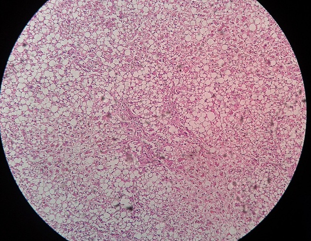
His hemoglobin was 8 g/dL and RBCs were microcytic hypochromic. Endoscopic biopsy revealed a few curved organisms and infiltration by neutrophils in the mucosa (fig. below). Lymphoid follicle formation was also found.
Tasks:
- What is the differential diagnosis of the above mentioned sign/symptoms?
- What is etiology and pathogenesis of the disease?
- Explain in detail the morphological changes seen in the gastric biopsy.
- What are special techniques used by the pathologists to reach exact type of the diagnosis?
- Describe the possible complications caused by this disease.
 howMed Know Yourself


