Necrosis
It is a spectrum of morphological changes that follow cell death in living tissues, resulting from progressive degradative action of enzymes on injured tissues.
Caseous Necrosis
The term caseous is derived from the grass appearance of the area of necrosis, i.e. white and cheesy appearance. Caseous necrosis is encountered most often in tuberculous infection. It is, in fact, a distinctive from of coagulative necrosis.
Mechanism
Macrophages
They have phagocytic activity for ingestion of microorganisms. There are certain organisms which are not killed by macrophages, e.g. mycobacterium tuberculosis. Macrophages engulf these organisms, retain them and present them to T lymphocytes.
T lymphocytes
They become activated and produce certain cytokines such as interferon gamma and interleukin 2. These cytokines activate other macrophages which are then transformed into epithelial cells. Epitheloid cells fuse together and form multi-nucleated giant cells. Some macrophages themselves die in this process and contribute to large areas of necrosis by release of their lysosomal enzymes. This process is known as cell mediated immunity or chronic granulomatous inflammation.
Epitheloid Cells
They have pale pink granular cytoplasm and differ from macrophages in that their nucleus is less dense, ovalent shape and vesicular in appearance. They have little phagocytic activity but have secretory function.
Multi-Nucleated Giant Cells
Giant cell is formed by the fusion of epitheloid cells and contain twenty or more nuclei. In case of tuberculous infection, Langhan giant cells are seen with nuclei arranged peripherally in shape of horse shoe. They may be seen in other granulomatous inflammations.

Morphology
Gross
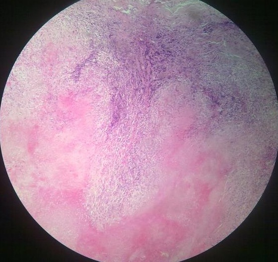
On gross inspection, dead tissue is seen as whitish and soft area with cream cheese consistency.
Microscopic Appearance
In the area of necrosis, normal architecture of tissues is completely obliterated. The necrotic area appears as eosinophilic, amorphous, granular debri composed of necrotic cells. Inflammatory border around the necrotic debri is known as granulomatous reaction. It consists of outermost rim of fibroblasts. Internal to this layer are T lymphocytes, then macrophages and epitheloid cells and occasional plasma cells. Langhan giant cells may also be present.
Other Types of Giant Cells
Foreign Body Giant Cells
There is central group of randomly arranged nuclei. They are found in inflammatory reaction around an old suture.
Tumor Giant Cells
They are present in association with tumor. No morphological nuclear arrangement is present.
Aschoff Giant Cells
They are present in rheumatic carditis. It is just an aggregate of cells and does not show any characteristic appearance.
Want a clearer concept, also see all images on Caseous Necrosis
 howMed Know Yourself


It really helps me a lot.