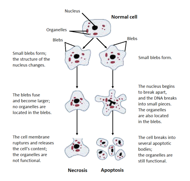
Necrosis is the denaturation of proteins & enzymatic digestion. It is irreversible local cell death and cellular dissolution in living tissue. It is auto digestion and lysis. Release of catalytic enzymes from lysosomes takes place resulting in autolysis/hydrolysis.
When severe tissue damage occurs, the metabolism stops, structure is destroyed and function is lost. E.g. histological evidence of myocardial necrosis is seen after 4-6 hrs.

Cytoplasmic changes
Cytoplasmic changes in necrosis include:
• Increased eosinophilia due to decreased cytoplasmic RNA & denatured cytoplasmic proteins, aggregates of fluffy material are also found.
• Increased glassy appearance due decreased glycogen.
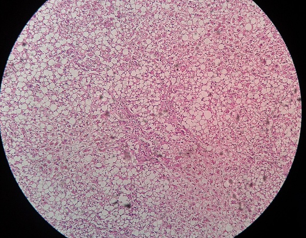
• Vacuolated cytoplasm after digestion.
• Myelin figures are whorled phospholipid masses derived from cell membrane.
• Dystrophic calcification of residual fatty acids.
Nuclear changes patterns
1. Karyolysis: decreased basophilia of chromatin
2. Pyknosis: nuclear shrinkage with increased basophilia
3. Karyorrhesis: fragmentation of pyknotic nuclei
Morphological changes in necrosis are due to:
• Enzymatic digestion of cells
• Denaturation of protein
Types of Necrosis
Steps/Sequel of necrosis
• Autolysis
• Phagocytosis
• Organization /fibrous repair
• Dystrophic calcification
Consequences of Necrosis
• Acute or chronic inflammation
• Immunological reactions to sub cellular components released by dead tissue or self-antigens altered by denaturation.
 howMed Know Yourself


