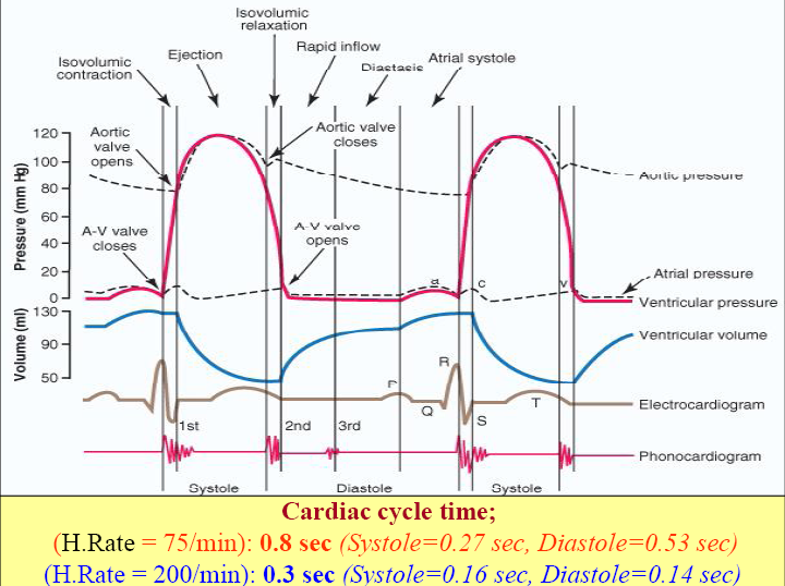
Cardiac events appearing from the beginning of one heart beat to the beginning of next heart beat and repeating themselves successively.
• Cardiac cycle time (H.Rate = 75/min) : 0.8 sec
Systole : 0.27 sec
Diastole: 0.53 sec
• Systole : Contraction and emptying
Muscle stimulated by action potential and contracting
• Diastole : Relaxation and filling
Muscle reestablishing Na+/K+/Ca++ gradient and is relaxing
Atria and ventricles go through separate cycles of
systole and diastole.
Atria contract prior to the ventricular contraction i.e.
when atria contract ventricles relax and vice versa.
Each ventricle expels same volume of blood/beat.
Systole:
1) Isovolumic contraction
A-V valves close;
because (ventricular press > atrial press)
2) Aortic valve opens
3) Ejection phase
4) Aortic valve closes
Diastole
1) Isovolumic relaxation
2) A-V valves open
3) Rapid inflow
4) Diastasis – slow flow into ventricle
5) Atrial systole – extra blood in and this just
follows P wave. Accounts for 25% of filling
Atrial Pressure/Jugular Venous Pressure Waves
Atrial pressure waves:
a-wave = atrial contraction
c-wave = ventricular contraction
(A-V valves bulge)
v-wave = flow of blood into atria
x- descent = falling Rt atrial pressure due to atrial diastole
y- descent = falling Rt atrial pressure during ventricular filling
Ejection Fraction
• End diastolic volume = 120 ml
• End systolic volume = 50 ml
• Ejection volume (stroke volume) = 70 ml
• Ejection fraction = 70ml/120ml = 58% (normally 60%)
• If heart rate (HR) is 70 beats/minute, what is cardiac
output?
• Cardiac output = HR x stroke volume
= 70/min. x 70 ml
= 4900ml/min.
• If HR =100, end diastolic volume = 180 ml,
end systolic vol. = 20 ml, what is cardiac output?
• C.O. = 100/min. x 160 ml = 16,000 ml/min.
Aortic Pressure Curve
• Aortic pressure starts increasing during systole
after the aortic valve opens.
• Aortic pressure decreases toward the end of the
ejection phase.
• After the aortic valve closes, an incisura occurs
because of sudden cessation of back-flow toward
left ventricle.
• Aortic pressure decreases slowly during diastole
because of the elasticity of the aorta.
Valvular Function
• To prevent back-flow.
• Chordae tendineae are attached to A-V valves.
• Papillary muscle, attached to chordae tendineae, contract during systole and help prevent back-flow.
• Because of smaller opening, velocity through aortic and pulmonary valves exceeds that through the A-V valves.
Events in Cardiac cycle (Left ventricle)
Systole : 0.27 Sec or 0.3 sec
LV : 120 mmHg
RV : 25 mmHg
1. Onset of ventricular contraction.
2. A-V valves closure.
3. Isovolumetric ventricular contraction.
(LV = 60 msec, RV = 15 msec)
4. Semilunar valves open.
5. Ventricular ejection. (LV= 200 msec, RV= 270 m sec)
a. Rapid ejection 1/3rd (70%)
b. Slow ejection 2/3rd (30%)
ESV = 65 ml.
6. Semilunar valves close.
Events in Cardiac cycle (Left ventricle)
Diastole: 0.53 sec or 0.5 sec
7. Isovolumetric relaxation (100 msec)
8. A.V valves open
9. Ventricular filling (500 msec)
a. Rapid filling 60-65%
b. Slow filling (Diastasis) 5-10%
c. Rapid filling again (Atrial systole) 20-25%
‘Atrial kick’
• EDV = 135 ml
• Atrial Systole:
a. RA = 4-6 mmHg
b. LA = 7-8 mmHg
 howMed Know Yourself

