Stress beyond the adaptive limit of the cell results in cell injury. Cell injury may be:
a. Reversible injury: stimulus is mild & transient & cells recover their lost functions.
b. Irreversible injury: stimulus is persistent & severe enough, leading to cell death.
Mechanism of cell injury
Injury to cell depends upon:
• Dose of injury
• State of target cell
• Susceptible cell components: mitochondria, cell membrane, rough endoplasmic reticulum, DNA.
Morphology of Reversible Injury
Light Microscopy
In case of reversible injury, following changes are observed under light microscopy:
• Cellular swelling
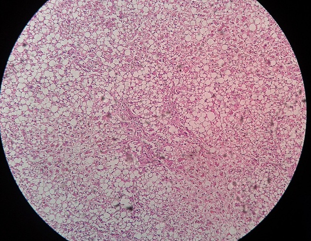
• Hydropic change / vacuolar degeneration.
• Failure of ionic pump of membrane
• Small clear vacuoles formed by pinched-off endoplasmic reticulum segments
• Increased eosinophilic staining of cytoplasm.
• Fatty change in hypoxic & toxic injuries
• Lipid vacuoles in cytoplasm
• Hepatocytes & myocardial cells
Electron Microscopy
Under electron microscopy, we find:
• Plasma membrane blebbing, blunting and loss of microvilli.
• Mitochondrial swelling & small amorphous densities.
• Endoplasmic reticulum dilatation and myelin figures
• Nuclear separation of granular & fibrillar elements
Morphology of Irreversible Cell Injury
• Increased swelling of organelles
• Disruption of lysosome
• Calcium deposits in mitochondria
• Disruption of membrane by phospholipase
Nuclear changes:
– Pyknosis (shrinkage with increased basophilia)
– Karyolysis
– Karyohexsis
– Anucleate cell
Want to clear your concepts, also see
 howMed Know Yourself


