Principle
The working principle of microscope is to show large image of those objects which cannot be seen with the naked eye.
Types of Microscope
The different types of microscope are:
- Light microscope/compound microscope
- Electron microscope
- Fluorescent microscope
- Dark field microscope
 Light/Compound Microscope
It is used in microbiology laboratory and uses yellow light which has a wavelength of 0.4 micrometers and a resolution of 0.2 micrometers i.e. half the wavelength of light is used.
Parts of a Compound Microscope
A compound binocular microscope consists of:
a. Eye Piece
It has a magnification of 10x and is set according to the pupils of the person using the microscope.
b. Objective Lenses
There are four objective lenses having power of 4x, 10x, 40x and 100x.
c. Stage
This is to place and fix the slide.
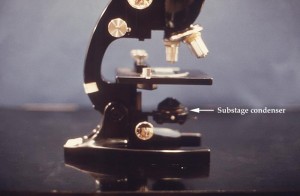
d. Condenser
It converges light from light source onto the slide.
e. Lamp
This is basically the source of light
f. Control Knobs
These are for fine and course adjustment.
How to Use a Microscope?
- Fix the slide on the stage
- Adjust light with the help of condenser
- Start focusing from lowest magnification using the coarse and fine adjustment knobs, once you focus it, move gradually from lower power objective lens (10x) to oil immersion lens (100x). Always use cedar wood oil to visualize the object under 100x objective.
- Always switch off the microscope when not in use.