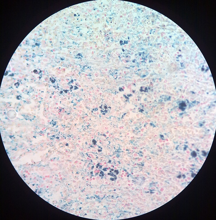
Hemosiderin is an endogenous blood derived golden yellow to brown granular or crystalline pigment. Other endogenous pigments are:
- Lipofuscin
- Melanin
- Bilirubin
- Porphyrin
Hemosiderosis is accumulation of hemosiderin in the cells due to stress of iron. The excess of iron may be local or systemic. The systemic excess is caused by:
- Excess intake of iron
- Repeated blood transfusion
- Hemolytic anemia

In systemic excess, iron is stored in mononuclear phagocytic cells of bone marrow, spleen, liver and lymph nodes. Iron is also taken up by scattered macrophages in different organs like skin, pancreas and kidney. When there is more accumulation of iron, it is stored in parenchymal cells of heart, liver and pancreas, leading to damage to these cells. This excess accumulation causes parenchymal damage in the viscera and condition is called hemochromatosis. In hemochromatosis, there may be damage to myocardium leading to heart failure, damage to liver leads to fibrosis and damage to pancreas leads to diabetes.
Want a clearer concept, also see all images on Hemosiderosis
 howMed Know Yourself


