Hydropic change or cellular swelling or vacuolar degeneration is one of the factors of reversible cell injury, which can be appreciated under light microscope.
Types of Reversible Cell Injury
- Hydropic change
- Fatty change
Types of Irreversible Cell Injury
It occurs if injurious stimulus is persistant.
- Necrosis
- Apoptosis
Pathogenesis
Cellular swelling is first manifestation of almost all forms of injury to cell. Two major stimuli which can lead to hydropic change are:
- Ischemia
- Chemical aging
Hydropic Change due to Ischemia
- Loss of blood supply leads to decreased oxygen tension inside cell and results in ATP depletion. There is also loss of oxidative phosphorylation causing decreased ATP generation and failure of Na+K+ pump. This leads to increased intracellular sodium and water along with increased extracellular potassium, leading to cellular swelling.
- Activation of anaerobic pathway causes accumulation of catabolites (phosphates and lactates) causing increased osmotic load and cellular swelling.
Hydropic Change due to Chemical Agents
a. Directly Acting Chemicals
They cause increased cell membrane injury leading to cellular injury e.g. cyanide poisoning, mercuric chloride, antineoplastic drugs and antibiotics.
b. Indirectly Acting Chemicals
They release highly toxic and reactive free radicals causing lipid peroxidation and cell membrane damage with increased influx of sodium and water causing cellular swelling e.g. carbon tetrachloride poisoning.

Morphology
Gross
Damaged organ e.g. kidney increases in weight, becomes swollen and pale in colour.
Microscopic
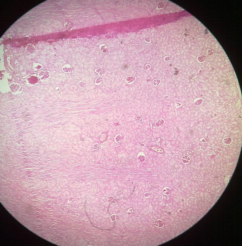
- It mainly involves renal tubules. Glomeruli are usually spared.
- Tubular cells become swollen and lightly stained.
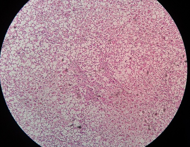
- Small, clear vacuoles are seen in the cytoplasm, which may be water vacuoles or represent distended endoplasmic reticulum.
- There may be granular appearance of cells due to presence of swollen mitochondria.
- Due to swelling of cells, lumen of renal tubules becomes narrow or completely obliterated. Swollen tubules compress the microvasculature present between them.
Ultramicroscopic
Electron microscopic changes of reversible cell injury include:
- Plasma membrane alterations such as blebbing, blunting and loss of microvilli.
- Mitochondrial changes include swelling and appearance of small, amorphous densities
- Dilation of endoplasmic reticulum and intra-cytoplasmic myelin figures may be present.
- Nuclear alteration with dis aggregation of granular and fibrillar elements.
Cells may be separated from basement membrane, nuclei pushed to one side, cells may become anucleated and there is no space between the tubules in very severe forms.
 howMed Know Yourself


