Hypertrophy is the increased size of cell, due to increased synthesis of structural components. It may be because of increased functional demand (e.g. increased workload for striated muscle) or increased stimulation by hormones & growth factors (uterine smooth muscle in pregnancy).
Hypertrophy beyond adaptive capacity leads to degenerative changes and organ failure. Common examples of hypertrophy include:
• Skeletal muscle in athletes
• Smooth muscle in gravid uterus
• Cardiac muscle in hypertension
• Remaining kidney after partial nephrectomy.

Mechanism of Cardiac Muscle Hypertrophy
In case of cardiac muscle hypertrophy, mechanical sensor is triggered by increased workload. Growth factors involved include TGF-β, IGF-1, FGF, α adrenergic agonists, Endothelin-1 and Angiotensin-II. Phosphoinositide 3-kinase/AKT pathway is activated through exercise or is physiologic. G-protein coupled receptor activation is pathologic. Switching of adult contractile protein to fetal form may occur (α isoform myosin to β isoform). Stress beyond the limit of hypertrophy results in regression, with loss of myofibrillar contractile proteins or cell death.
Mechanism of Hypertrophy of Endoplasmic Reticulum
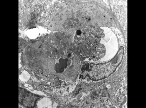
Endoplasmic reticulum is a subcellular organelle. Barbiturates induce hypertrophy of smooth endoplasmic reticulum occurs in hepatocytes, leading to increased enzyme cytochrome P-450 mixed function oxidase.
Examples of hypertrophy
Hypertrophy may be congenital or acquired.
Muscular hypertrophy
Skeletal muscles in response to strength training (known as muscle hypertrophy). Depending on the type of training, the hypertrophy can occur through increased sarcoplasmic volume or increased contractile proteins.
Ventricular hypertrophy
Ventricular hypertrophy is the increase in size of the ventricles of the heart. Changes can be beneficial if they occur in response to aerobic or anaerobic exercise, but ventricular hypertrophy is generally associated with pathological changes due to high blood pressure or valvular heart disease states.
 howMed Know Yourself


