Pathological calcification is abnormal deposition of calcium salts together with small amounts of magnesium, iron and other minerals. It is of two types:
- Dystrophic Calcification
- Metastatic Calcification
Dystrophic Calcification
It is deposition of calcium salts in dead or dying tissues without any derangement of calcium metabolism. It is commonly seen in areas of necrosis, caseous and enzymatic fat necrosis are common. The common sites of occurrence are:
- Atheromas
- Damaged heart valves
- Tuberculos lymph nodes
- Papillary carcinoma of thyroid and ovaries
- Meningiomas
- May also be seen in dead parasites and fetus
Metastatic Calcification
It occurs in normal tissues, in presence of hypercalcemia. The causes of hypercalcemia are:
a. Increased secretion of parathyroid
- Parathyroid adenoma
- Parathyroid hyperplasia
- Parathyroid carcinoma
- Ectopic secretion of parathyroid hormone related protein e.g. by squamous cell carcinoma of lung, breast carcinoma, renal cell carcinoma.
- Adult T cell leukemia or lymphoma and ovarian carcinoma.
b. Destruction of Bone Tissue
- Primary tumor of bone marrow e.g. leukemia and multiple myeloma
- Skeletal metastasis
- Increased bone turn over e.g. in Paget’s disease
- Immobilization
c. Renal Failure
Phosphate retention leads to secondary hyperparathyroidism
d. Uncommon Causes
- Aluminium intoxication e.g. with chronic renal dialysis
- Milk alkali syndrome
Metastatic calcification may occur throughout body but principally affected sites are:
- Gastric mucosa
- Kidney
- Lungs
- Systemic arteries
- Pulmonary veins
Morphology
Gross
Whatever the site of deposition, calcium salts appear as fine white colored granules or clumps, which are gritty in consistency.
Microscopy
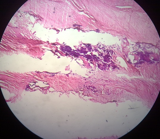
Calcium salts can be visualized as basophilic, amorphous granules or clumps, which are present intracellularly or extracellularly. As time passes, heterotrophic bone formation may be seen in foci of calcification.
In asbestosis, calcium and iron salts gather about large cylinder spicules of asbestos in lungs, creating dumb bell forms.
The calcified deposits may have concentric lamellar appearance, known as Psammoma bodies, this is found in some types of papillary carcinoma of thyroid.
Want a clearer concept, also see all images on Calcification
 howMed Know Yourself


